3FDE

Mouse UHRF1 SRA domain bound with hemi-methylated CpG DNA, crystal structure in space group C222(1) at 1.4 A resolution


Experimental Data Snapshot

  • Method: X-RAY DIFFRACTION
  • Resolution: 1.41 Å
  • R-Value Free: 0.186 
  • R-Value Work: 0.149 
  • R-Value Observed: 0.151 

wwPDB Validation 

Currently 3FDE does not have a validation slider image.


This is version 1.2 of the entry. See complete history


Literature

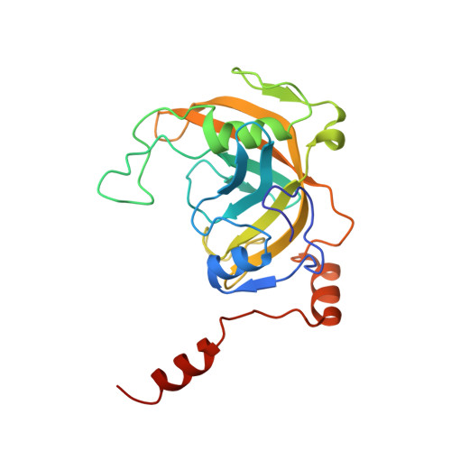
UHRF1, a modular multi-domain protein, regulates replication-coupled crosstalk between DNA methylation and histone modifications.

Hashimoto, H.Horton, J.R.Zhang, X.Cheng, X.

(2009) Epigenetics 4: 8-14

  • DOI: https://doi.org/10.4161/epi.4.1.7370
  • Primary Citation of Related Structures:  
    3F8I, 3F8J, 3FDE

  • PubMed Abstract: 

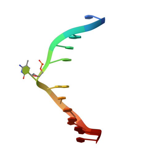
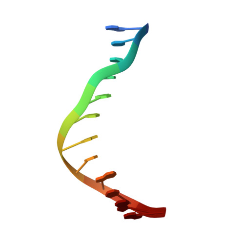
    Cytosine methylation in DNA is a major epigenetic signal, and plays a central role in propagating chromatin status during cell division. However the mechanistic links between DNA methylation and histone methylation are poorly understood. A multi-domain protein UHRF1 (ubiquitin-like, containing PHD and RING finger domains 1) is required for DNA CpG maintenance methylation at replication forks, and mouse UHRF1-null cells show enhanced susceptibility to DNA replication arrest and DNA damaging agents. Recent data demonstrated that the SET and RING associated (SRA) domain of UHRF1 binds hemimethylated CpG and flips 5-methylcytosine out of the DNA helix, whereas its tandom tudor domain and PHD domain bind the tail of histone H3 in a highly methylation sensitive manner. We hypothesize that UHRF1 brings the two components (histones and DNA) carrying appropriate markers (on the tails of H3 and hemimethylated CpG sites) ready to be assembled into a nucleosome after replication.


  • Organizational Affiliation

    Department of Biochemistry, Emory University School of Medicine, Atlanta, Georgia 30322, USA.


Macromolecules

Find similar proteins by:  (by identity cutoff)  |  3D Structure
Entity ID: 1
MoleculeChains Sequence LengthOrganismDetailsImage
E3 ubiquitin-protein ligase UHRF1
A, B
212Mus musculusMutation(s): 0 
Gene Names: Np95Uhrf1
EC: 6.3.2
UniProt & NIH Common Fund Data Resources
Find proteins for Q8VDF2 (Mus musculus)
Explore Q8VDF2 
Go to UniProtKB:  Q8VDF2
IMPC:  MGI:1338889
Entity Groups  
Sequence Clusters30% Identity50% Identity70% Identity90% Identity95% Identity100% Identity
UniProt GroupQ8VDF2
Sequence Annotations
Expand
  • Reference Sequence

Find similar nucleic acids by:  Sequence   |   3D Structure  

Entity ID: 2
MoleculeChains LengthOrganismImage
5'-D(*CP*CP*AP*TP*GP*(5CM)P*GP*CP*TP*GP*AP*C)-3'C [auth D],
E [auth C]
12N/A
Sequence Annotations
Expand
  • Reference Sequence

Find similar nucleic acids by:  Sequence   |   3D Structure  

Entity ID: 3
MoleculeChains LengthOrganismImage
5'-D(*GP*TP*CP*AP*GP*CP*GP*CP*AP*TP*GP*G)-3'D [auth E],
F
12N/A
Sequence Annotations
Expand
  • Reference Sequence
Small Molecules
Ligands 3 Unique
IDChains Name / Formula / InChI Key2D Diagram3D Interactions
EDO
Query on EDO

Download Ideal Coordinates CCD File 
BA [auth D]
CA [auth E]
DA [auth E]
FA [auth C]
G [auth A]
BA [auth D],
CA [auth E],
DA [auth E],
FA [auth C],
G [auth A],
H [auth A],
I [auth A],
J [auth A],
JA [auth F],
K [auth A],
KA [auth F],
L [auth A],
LA [auth F],
M [auth A],
MA [auth F],
N [auth A],
NA [auth F],
R [auth B],
S [auth B],
T [auth B],
U [auth B],
V [auth B]
1,2-ETHANEDIOL
C2 H6 O2
LYCAIKOWRPUZTN-UHFFFAOYSA-N
NA
Query on NA

Download Ideal Coordinates CCD File 
EA [auth E],
O [auth A],
OA [auth F],
W [auth B]
SODIUM ION
Na
FKNQFGJONOIPTF-UHFFFAOYSA-N
UNL
Query on UNL

Download Ideal Coordinates CCD File 
AA [auth B]
GA [auth C]
HA [auth C]
IA [auth C]
P [auth A]
AA [auth B],
GA [auth C],
HA [auth C],
IA [auth C],
P [auth A],
Q [auth A],
X [auth B],
Y [auth B],
Z [auth B]
Unknown ligand
FKNQFGJONOIPTF-UHFFFAOYSA-N
Experimental Data & Validation

Experimental Data

  • Method: X-RAY DIFFRACTION
  • Resolution: 1.41 Å
  • R-Value Free: 0.186 
  • R-Value Work: 0.149 
  • R-Value Observed: 0.151 
  • Space Group: C 2 2 21
Unit Cell:
Length ( Å )Angle ( ˚ )
a = 84.678α = 90
b = 103.691β = 90
c = 149.838γ = 90
Software Package:
Software NamePurpose
HKL-2000data collection
PHASERphasing
REFMACrefinement
HKL-2000data reduction
HKL-2000data scaling

Structure Validation

Currently 3FDE does not have a validation slider image.


Entry History 

Deposition Data

Revision History  (Full details and data files)

  • Version 1.0: 2009-01-06
    Type: Initial release
  • Version 1.1: 2011-07-13
    Changes: Version format compliance
  • Version 1.2: 2023-12-27
    Changes: Advisory, Data collection, Database references, Derived calculations