3EDV

Crystal Structure of Repeats 14-16 of Beta2-Spectrin


Experimental Data Snapshot

  • Method: X-RAY DIFFRACTION
  • Resolution: 1.95 Å
  • R-Value Free: 0.250 
  • R-Value Work: 0.192 
  • R-Value Observed: 0.195 

wwPDB Validation   3D Report Full Report


This is version 1.2 of the entry. See complete history


Literature

Localization and Structure of the Ankyrin-binding Site on beta2-Spectrin

Davis, L.Abdi, K.Machius, M.Brautigam, C.Tomchick, D.R.Bennett, V.Michaely, P.

(2009) J Biol Chem 284: 6982-6987

  • DOI: https://doi.org/10.1074/jbc.M809245200
  • Primary Citation of Related Structures:  
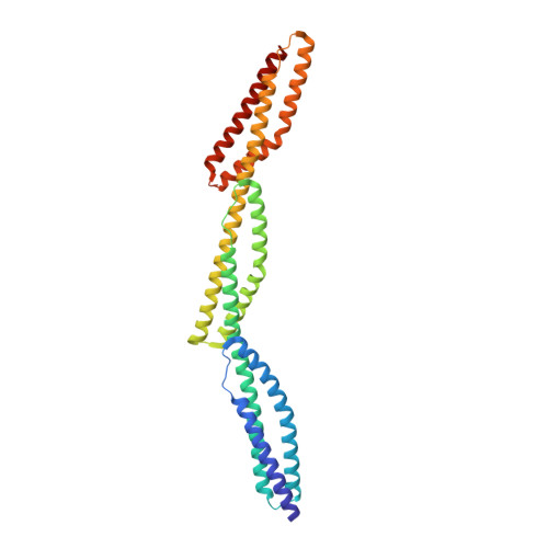
    3EDV

  • PubMed Abstract: 

    Spectrins are tetrameric actin-cross-linking proteins that form an elastic network, termed the membrane skeleton, on the cytoplasmic surface of cellular membranes. At the plasma membrane, the membrane skeleton provides essential support, preventing loss of membrane material to environmental shear stresses. The skeleton also controls the location, abundance, and activity of membrane proteins that are critical to cell and tissue function. The ability of the skeleton to modulate membrane stability and function requires adaptor proteins that bind the skeleton to membranes. The principal adaptors are the ankyrin proteins, which bind to the beta-subunit of spectrin and to the cytoplasmic domains of numerous integral membrane proteins. Here, we present the crystal structure of the ankyrin-binding domain of human beta2-spectrin at 1.95 A resolution together with mutagenesis data identifying the binding surface for ankyrins on beta2-spectrin.


  • Organizational Affiliation

    Department of Cell Biology and Howard Hughes Medical Institute, Duke University Medical Center, Durham, North Carolina 27710, USA.


Macromolecules
Find similar proteins by:  (by identity cutoff)  |  3D Structure
Entity ID: 1
MoleculeChains Sequence LengthOrganismDetailsImage
Spectrin beta chain, brain 1
A, B
323Homo sapiensMutation(s): 0 
Gene Names: SPTBN1SPTB2
UniProt & NIH Common Fund Data Resources
Find proteins for Q01082 (Homo sapiens)
Explore Q01082 
Go to UniProtKB:  Q01082
PHAROS:  Q01082
GTEx:  ENSG00000115306 
Entity Groups  
Sequence Clusters30% Identity50% Identity70% Identity90% Identity95% Identity100% Identity
UniProt GroupQ01082
Sequence Annotations
Expand
  • Reference Sequence
Small Molecules
Ligands 1 Unique
IDChains Name / Formula / InChI Key2D Diagram3D Interactions
MG
Query on MG

Download Ideal Coordinates CCD File 
C [auth A]
D [auth A]
E [auth A]
F [auth A]
G [auth A]
C [auth A],
D [auth A],
E [auth A],
F [auth A],
G [auth A],
H [auth A],
I [auth B],
J [auth B],
K [auth B],
L [auth B],
M [auth B],
N [auth B],
O [auth B]
MAGNESIUM ION
Mg
JLVVSXFLKOJNIY-UHFFFAOYSA-N
Experimental Data & Validation

Experimental Data

  • Method: X-RAY DIFFRACTION
  • Resolution: 1.95 Å
  • R-Value Free: 0.250 
  • R-Value Work: 0.192 
  • R-Value Observed: 0.195 
  • Space Group: P 21 21 21
Unit Cell:
Length ( Å )Angle ( ˚ )
a = 51.335α = 90
b = 56.415β = 90
c = 261.611γ = 90
Software Package:
Software NamePurpose
PHENIXrefinement
HKL-3000data collection
HKL-3000data reduction
HKL-3000data scaling
SHELXDphasing

Structure Validation

View Full Validation Report


Entry History 

Deposition Data

Revision History  (Full details and data files)

  • Version 1.0: 2009-01-20
    Type: Initial release
  • Version 1.1: 2011-07-13
    Changes: Version format compliance
  • Version 1.2: 2024-02-21
    Changes: Data collection, Database references, Derived calculations