


3DDW
Crystal structure of glycogen phosphorylase complexed with an anthranilimide based inhibitor GSK055
- PDB DOI: https://doi.org/10.2210/pdb3DDW/pdb
- Classification: TRANSFERASE
- Organism(s): Homo sapiens
- Expression System: Escherichia coli
- Mutation(s): No
- Deposited: 2008-06-06 Released: 2009-01-27
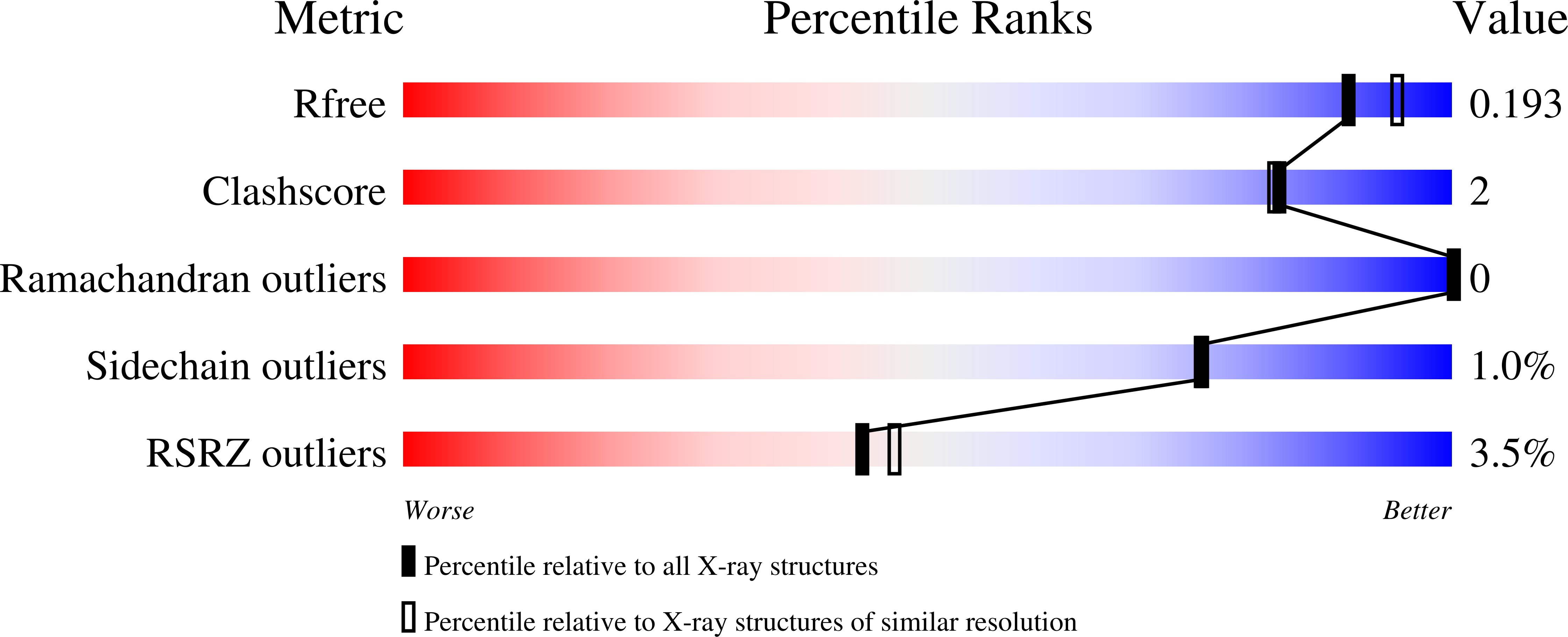
Experimental Data Snapshot
- Method: X-RAY DIFFRACTION
- Resolution: 1.90 Å
- R-Value Free: 0.186
- R-Value Work: 0.153
- R-Value Observed: 0.154
This is version 1.3 of the entry. See complete history.

Macromolecules
Find similar proteins by:
(by identity cutoff) | 3D Structure
Entity ID: 1 | |||||
|---|---|---|---|---|---|
| Molecule | Chains | Sequence Length | Organism | Details | Image |
| Glycogen phosphorylase, liver form | 848 | Homo sapiens | Mutation(s): 0 Gene Names: PYGL EC: 2.4.1.1 | 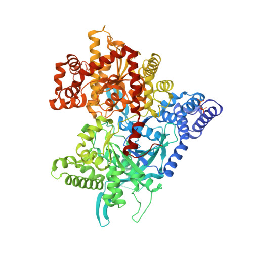 | |
UniProt & NIH Common Fund Data Resources | |||||
Find proteins for P06737 (Homo sapiens) Explore P06737 Go to UniProtKB: P06737 | |||||
PHAROS: P06737 GTEx: ENSG00000100504 | |||||
Entity Groups | |||||
| Sequence Clusters | 30% Identity50% Identity70% Identity90% Identity95% Identity100% Identity | ||||
| UniProt Group | P06737 | ||||
Sequence AnnotationsExpand | |||||
| |||||
Small Molecules
| Ligands 6 Unique | |||||
|---|---|---|---|---|---|
| ID | Chains | Name / Formula / InChI Key | 2D Diagram | 3D Interactions | |
| 055 Query on 055 | F [auth A], M [auth B] | (2S)-{[(3-{[(2-chloro-6-methylphenyl)carbamoyl]amino}naphthalen-2-yl)carbonyl]amino}(phenyl)ethanoic acid C27 H22 Cl N3 O4 KRIVDSIBMDCVLL-DEOSSOPVSA-N |  | ||
| PLP Query on PLP | D [auth A], I [auth B] | PYRIDOXAL-5'-PHOSPHATE C8 H10 N O6 P NGVDGCNFYWLIFO-UHFFFAOYSA-N |  | ||
| NBG Query on NBG | C [auth A], H [auth B] | N-acetyl-beta-D-glucopyranosylamine C8 H15 N O6 IBONACLSSOLHFU-JAJWTYFOSA-N |  | ||
| MES Query on MES | K [auth B], L [auth B] | 2-(N-MORPHOLINO)-ETHANESULFONIC ACID C6 H13 N O4 S SXGZJKUKBWWHRA-UHFFFAOYSA-N |  | ||
| CFF Query on CFF | E [auth A], J [auth B] | CAFFEINE C8 H10 N4 O2 RYYVLZVUVIJVGH-UHFFFAOYSA-N |  | ||
| MPD Query on MPD | G [auth A], N [auth B] | (4S)-2-METHYL-2,4-PENTANEDIOL C6 H14 O2 SVTBMSDMJJWYQN-YFKPBYRVSA-N |  | ||
| Modified Residues 1 Unique | |||||
|---|---|---|---|---|---|
| ID | Chains | Type | Formula | 2D Diagram | Parent |
| SEP Query on SEP | A, B | L-PEPTIDE LINKING | C3 H8 N O6 P |  | SER |
Experimental Data & Validation
Experimental Data
- Method: X-RAY DIFFRACTION
- Resolution: 1.90 Å
- R-Value Free: 0.186
- R-Value Work: 0.153
- R-Value Observed: 0.154
- Space Group: P 31
Unit Cell:
| Length ( Å ) | Angle ( ˚ ) |
|---|---|
| a = 124.368 | α = 90 |
| b = 124.368 | β = 90 |
| c = 123.621 | γ = 120 |
| Software Name | Purpose |
|---|---|
| REFMAC | refinement |
| ADSC | data collection |
| HKL-2000 | data reduction |
| HKL-2000 | data scaling |
| AMoRE | phasing |
Entry History
Deposition Data
- Released Date: 2009-01-27 Deposition Author(s): Nolte, R.T.
Revision History (Full details and data files)
- Version 1.0: 2009-01-27
Type: Initial release - Version 1.1: 2011-07-13
Changes: Non-polymer description, Version format compliance - Version 1.2: 2020-07-29
Type: Remediation
Reason: Carbohydrate remediation
Changes: Data collection, Database references, Derived calculations, Structure summary - Version 1.3: 2023-08-30
Changes: Data collection, Database references, Refinement description, Structure summary
