3D6A

Crystal structure of the 2H-phosphatase domain of Sts-2 in complex with tungstate.


Experimental Data Snapshot

  • Method: X-RAY DIFFRACTION
  • Resolution: 2.25 Å
  • R-Value Free: 0.257 
  • R-Value Work: 0.208 
  • R-Value Observed: 0.211 

wwPDB Validation   3D Report Full Report


This is version 1.3 of the entry. See complete history


Literature

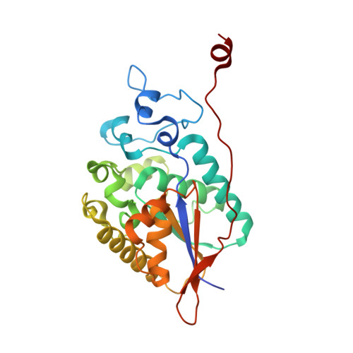
Structural and functional characterization of the 2H-phosphatase domain of Sts-2 reveals an acid-dependent phosphatase activity.

Chen, Y.Jakoncic, J.Carpino, N.Nassar, N.

(2009) Biochemistry 48: 1681-1690

  • DOI: https://doi.org/10.1021/bi802219n
  • Primary Citation of Related Structures:  
    3D4I, 3D6A, 3DB1

  • PubMed Abstract: 

    The suppressors of T cell receptor (TCR) signaling 1 and 2 (Sts-1 and -2, respectively) are multidomain proteins that negatively regulate the signaling of membrane-bound receptors, including TCR and the epidermal growth factor receptor (EGFR). Sts-1 was recently shown to be a new type of protein tyrosine phosphatase (PTP), with the phosphatase activity located within its C-terminal phosphoglycerate mutase (PGM) homology domain and key for the regulation of TCR signaling in T cells. The activity of the related Sts-2 enzyme is significantly less than that of Sts-1. Here we investigate the phosphatase activity of the PGM domain of Sts-2, Sts-2(PGM). The crystal structure of Sts-2(PGM) is remarkably similar to Sts-1(PGM), including conservation of all catalytic residues. Insight into mechanistic details is provided by the structures of the apo, tungstate-bound, and phosphate-bound enzyme. The active site shows stringent specificity, with the k(cat) optimum at pH 5.0 suggesting that Sts-2 might function as an acid-dependent phosphatase. Mutation of active site residues Gln372, Ala446, Glu481, Ser552, and Ser582 to their equivalents in Sts-1 increases the phosphatase activity of Sts-2(PGM) toward model substrates. Overall, our data demonstrate that Sts-2(PGM) adopts the conformation of an active phosphatase whose activity is fundamentally different from that of Sts-1 despite the strong structural homology. They also demonstrate that nonconserved active site residues are responsible for the difference in activity between the two isoforms. These differences reflect possible distinct physiological substrates.


  • Organizational Affiliation

    Department of Physiology and Biophysics, Stony Brook University, Stony Brook, New York 11794-8661, USA.


Macromolecules
Find similar proteins by:  (by identity cutoff)  |  3D Structure
Entity ID: 1
MoleculeChains Sequence LengthOrganismDetailsImage
Sts-2 protein
A, B, C, D
273Mus musculusMutation(s): 0 
Gene Names: Ubash3a
UniProt & NIH Common Fund Data Resources
Find proteins for Q3V3E1 (Mus musculus)
Explore Q3V3E1 
Go to UniProtKB:  Q3V3E1
IMPC:  MGI:1926074
Entity Groups  
Sequence Clusters30% Identity50% Identity70% Identity90% Identity95% Identity100% Identity
UniProt GroupQ3V3E1
Sequence Annotations
Expand
  • Reference Sequence
Small Molecules
Ligands 3 Unique
IDChains Name / Formula / InChI Key2D Diagram3D Interactions
W
Query on W

Download Ideal Coordinates CCD File 
J [auth A],
M [auth B],
Q [auth C],
U [auth D]
TUNGSTEN ION
W
FZFRVZDLZISPFJ-UHFFFAOYSA-N
MG
Query on MG

Download Ideal Coordinates CCD File 
E [auth A]
F [auth A]
G [auth A]
H [auth A]
K [auth B]
E [auth A],
F [auth A],
G [auth A],
H [auth A],
K [auth B],
N [auth C],
O [auth C],
R [auth D],
S [auth D]
MAGNESIUM ION
Mg
JLVVSXFLKOJNIY-UHFFFAOYSA-N
NA
Query on NA

Download Ideal Coordinates CCD File 
I [auth A],
L [auth B],
P [auth C],
T [auth D]
SODIUM ION
Na
FKNQFGJONOIPTF-UHFFFAOYSA-N
Experimental Data & Validation

Experimental Data

  • Method: X-RAY DIFFRACTION
  • Resolution: 2.25 Å
  • R-Value Free: 0.257 
  • R-Value Work: 0.208 
  • R-Value Observed: 0.211 
  • Space Group: P 21 21 21
Unit Cell:
Length ( Å )Angle ( ˚ )
a = 78.205α = 90
b = 116.665β = 90
c = 121.471γ = 90
Software Package:
Software NamePurpose
REFMACrefinement
HKL-2000data collection
HKL-2000data reduction
HKL-2000data scaling
CCP4phasing

Structure Validation

View Full Validation Report


Entry History 

Deposition Data

Revision History  (Full details and data files)

  • Version 1.0: 2009-03-03
    Type: Initial release
  • Version 1.1: 2011-07-13
    Changes: Advisory, Version format compliance
  • Version 1.2: 2023-08-30
    Changes: Data collection, Database references, Derived calculations, Refinement description
  • Version 1.3: 2024-03-13
    Changes: Source and taxonomy