3C7K

Molecular architecture of Galphao and the structural basis for RGS16-mediated deactivation


Experimental Data Snapshot

  • Method: X-RAY DIFFRACTION
  • Resolution: 2.90 Å
  • R-Value Free: 0.311 
  • R-Value Work: 0.250 

wwPDB Validation   3D Report Full Report


Ligand Structure Quality Assessment 


This is version 1.3 of the entry. See complete history


Literature

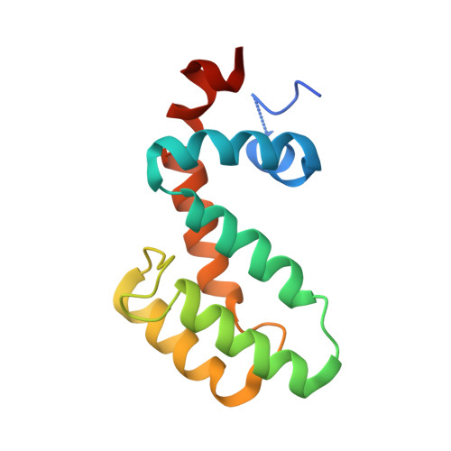
Molecular architecture of G{alpha}o and the structural basis for RGS16-mediated deactivation.

Slep, K.C.Kercher, M.A.Wieland, T.Chen, C.K.Simon, M.I.Sigler, P.B.

(2008) Proc Natl Acad Sci U S A 105: 6243-6248

  • DOI: https://doi.org/10.1073/pnas.0801569105
  • Primary Citation of Related Structures:  
    3C7K, 3C7L

  • PubMed Abstract: 

    Heterotrimeric G proteins relay extracellular cues from heptahelical transmembrane receptors to downstream effector molecules. Composed of an alpha subunit with intrinsic GTPase activity and a betagamma heterodimer, the trimeric complex dissociates upon receptor-mediated nucleotide exchange on the alpha subunit, enabling each component to engage downstream effector targets for either activation or inhibition as dictated in a particular pathway. To mitigate excessive effector engagement and concomitant signal transmission, the Galpha subunit's intrinsic activation timer (the rate of GTP hydrolysis) is regulated spatially and temporally by a class of GTPase accelerating proteins (GAPs) known as the regulator of G protein signaling (RGS) family. The array of G protein-coupled receptors, Galpha subunits, RGS proteins and downstream effectors in mammalian systems is vast. Understanding the molecular determinants of specificity is critical for a comprehensive mapping of the G protein system. Here, we present the 2.9 A crystal structure of the enigmatic, neuronal G protein Galpha(o) in the GTP hydrolytic transition state, complexed with RGS16. Comparison with the 1.89 A structure of apo-RGS16, also presented here, reveals plasticity upon Galpha(o) binding, the determinants for GAP activity, and the structurally unique features of Galpha(o) that likely distinguish it physiologically from other members of the larger Galpha(i) family, affording insight to receptor, GAP and effector specificity.


  • Organizational Affiliation

    Department of Biology, University of North Carolina, Chapel Hill, NC 27599, USA. kslep@bio.unc.edu


Macromolecules
Find similar proteins by:  (by identity cutoff)  |  3D Structure
Entity ID: 1
MoleculeChains Sequence LengthOrganismDetailsImage
Guanine nucleotide-binding protein G(o) subunit alpha
A, C
333Mus musculusMutation(s): 0 
Gene Names: Gnao1Gna0Gnao
UniProt & NIH Common Fund Data Resources
Find proteins for P18872 (Mus musculus)
Explore P18872 
Go to UniProtKB:  P18872
IMPC:  MGI:95775
Entity Groups  
Sequence Clusters30% Identity50% Identity70% Identity90% Identity95% Identity100% Identity
UniProt GroupP18872
Sequence Annotations
Expand
  • Reference Sequence
Find similar proteins by:  (by identity cutoff)  |  3D Structure
Entity ID: 2
MoleculeChains Sequence LengthOrganismDetailsImage
Regulator of G-protein signaling 16
B, D
129Mus musculusMutation(s): 0 
Gene Names: Rgs16Rgsr
UniProt
Find proteins for P97428 (Mus musculus)
Explore P97428 
Go to UniProtKB:  P97428
Entity Groups  
Sequence Clusters30% Identity50% Identity70% Identity90% Identity95% Identity100% Identity
UniProt GroupP97428
Sequence Annotations
Expand
  • Reference Sequence
Experimental Data & Validation

Experimental Data

  • Method: X-RAY DIFFRACTION
  • Resolution: 2.90 Å
  • R-Value Free: 0.311 
  • R-Value Work: 0.250 
  • Space Group: P 32 2 1
Unit Cell:
Length ( Å )Angle ( ˚ )
a = 96.357α = 90
b = 96.357β = 90
c = 235.61γ = 120
Software Package:
Software NamePurpose
DENZOdata reduction
SCALEPACKdata scaling
CNSrefinement
PDB_EXTRACTdata extraction
CNSphasing

Structure Validation

View Full Validation Report


Ligand Structure Quality Assessment 


Entry History 

Deposition Data

Revision History  (Full details and data files)

  • Version 1.0: 2008-05-06
    Type: Initial release
  • Version 1.1: 2011-07-13
    Changes: Version format compliance
  • Version 1.2: 2017-10-25
    Changes: Refinement description
  • Version 1.3: 2023-08-30
    Changes: Data collection, Database references, Derived calculations, Refinement description