3C4V

Structure of the retaining glycosyltransferase MshA:The first step in mycothiol biosynthesis. Organism: Corynebacterium glutamicum : Complex with UDP and 1L-INS-1-P.


Experimental Data Snapshot

  • Method: X-RAY DIFFRACTION
  • Resolution: 2.60 Å
  • R-Value Free: 0.213 
  • R-Value Work: 0.190 
  • R-Value Observed: 0.191 

wwPDB Validation   3D Report Full Report


Ligand Structure Quality Assessment 


This is version 1.2 of the entry. See complete history


Literature

Structural and Enzymatic Analysis of MshA from Corynebacterium glutamicum: SUBSTRATE-ASSISTED CATALYSIS

Vetting, M.W.Frantom, P.A.Blanchard, J.S.

(2008) J Biol Chem 283: 15834-15844

  • DOI: https://doi.org/10.1074/jbc.M801017200
  • Primary Citation of Related Structures:  
    3C48, 3C4Q, 3C4V

  • PubMed Abstract: 

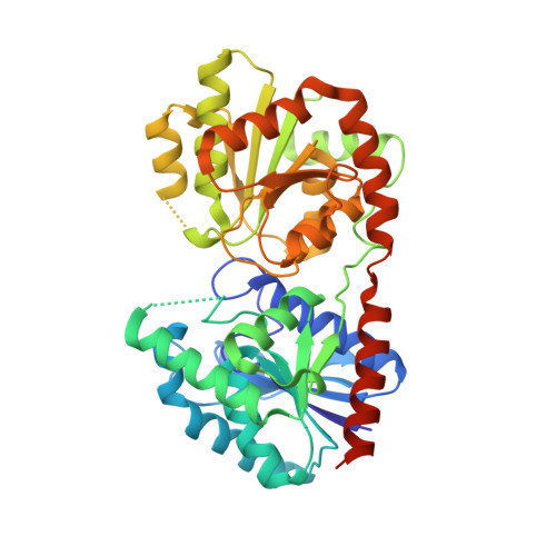
    The glycosyltransferase termed MshA catalyzes the transfer of N-acetylglucosamine from UDP-N-acetylglucosamine to 1-L-myo-inositol-1-phosphate in the first committed step of mycothiol biosynthesis. The structure of MshA from Corynebacterium glutamicum was determined both in the absence of substrates and in a complex with UDP and 1-L-myo-inositol-1-phosphate. MshA belongs to the GT-B structural family whose members have a two-domain structure with both domains exhibiting a Rossman-type fold. Binding of the donor sugar to the C-terminal domain produces a 97 degrees rotational reorientation of the N-terminal domain relative to the C-terminal domain, clamping down on UDP and generating the binding site for 1-L-myo-inositol-1-phosphate. The structure highlights the residues important in binding of UDP-N-acetylglucosamine and 1-L-myo-inositol-1-phosphate. Molecular models of the ternary complex suggest a mechanism in which the beta-phosphate of the substrate, UDP-N-acetylglucosamine, promotes the nucleophilic attack of the 3-hydroxyl group of 1-L-myo-inositol-1-phosphate while at the same time promoting the cleavage of the sugar nucleotide bond.


  • Organizational Affiliation

    Department of Biochemistry, Albert Einstein College of Medicine, Bronx, New York 10461, USA. vetting@aecom.yu.edu


Macromolecules
Find similar proteins by:  (by identity cutoff)  |  3D Structure
Entity ID: 1
MoleculeChains Sequence LengthOrganismDetailsImage
Predicted glycosyltransferases
A, B
426Corynebacterium glutamicumMutation(s): 0 
EC: 2.4.1
UniProt
Find proteins for Q8NTA6 (Corynebacterium glutamicum (strain ATCC 13032 / DSM 20300 / BCRC 11384 / JCM 1318 / LMG 3730 / NCIMB 10025))
Explore Q8NTA6 
Go to UniProtKB:  Q8NTA6
Entity Groups  
Sequence Clusters30% Identity50% Identity70% Identity90% Identity95% Identity100% Identity
UniProt GroupQ8NTA6
Sequence Annotations
Expand
  • Reference Sequence
Experimental Data & Validation

Experimental Data

  • Method: X-RAY DIFFRACTION
  • Resolution: 2.60 Å
  • R-Value Free: 0.213 
  • R-Value Work: 0.190 
  • R-Value Observed: 0.191 
  • Space Group: I 4 2 2
Unit Cell:
Length ( Å )Angle ( ˚ )
a = 223.94α = 90
b = 223.94β = 90
c = 125.005γ = 90
Software Package:
Software NamePurpose
REFMACrefinement
CBASSdata collection
MOSFLMdata reduction
SCALAdata scaling
AMoREphasing

Structure Validation

View Full Validation Report


Ligand Structure Quality Assessment 


Entry History 

Deposition Data

Revision History  (Full details and data files)

  • Version 1.0: 2008-04-01
    Type: Initial release
  • Version 1.1: 2011-07-13
    Changes: Version format compliance
  • Version 1.2: 2023-08-30
    Changes: Data collection, Database references, Derived calculations, Refinement description