2ZTA

X-RAY STRUCTURE OF THE GCN4 LEUCINE ZIPPER, A TWO-STRANDED, PARALLEL COILED COIL


Experimental Data Snapshot

  • Method: X-RAY DIFFRACTION
  • Resolution: 1.80 Å
  • R-Value Work: 0.179 

wwPDB Validation   3D Report Full Report


This is version 1.2 of the entry. See complete history


Literature

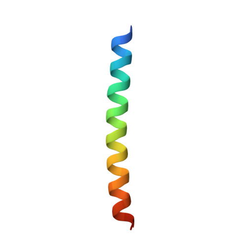
X-ray structure of the GCN4 leucine zipper, a two-stranded, parallel coiled coil.

O'Shea, E.K.Klemm, J.D.Kim, P.S.Alber, T.

(1991) Science 254: 539-544

  • DOI: https://doi.org/10.1126/science.1948029
  • Primary Citation of Related Structures:  
    2ZTA

  • PubMed Abstract: 

    The x-ray crystal structure of a peptide corresponding to the leucine zipper of the yeast transcriptional activator GCN4 has been determined at 1.8 angstrom resolution. The peptide forms a parallel, two-stranded coiled coil of alpha helices packed as in the "knobs-into-holes" model proposed by Crick in 1953. Contacts between the helices include ion pairs and an extensive hydrophobic interface that contains a distinctive hydrogen bond. The conserved leucines, like the residues in the alternate hydrophobic repeat, make side-to-side interactions (as in a handshake) in every other layer of the dimer interface. The crystal structure of the GCN4 leucine zipper suggests a key role for the leucine repeat, but also shows how other features of the coiled coil contribute to dimer formation.


  • Organizational Affiliation

    Howard Hughes Medical Institute, Cambridge, MA 02142.


Macromolecules
Find similar proteins by:  (by identity cutoff)  |  3D Structure
Entity ID: 1
MoleculeChains Sequence LengthOrganismDetailsImage
GCN4 LEUCINE ZIPPER
A, B
34Saccharomyces cerevisiaeMutation(s): 0 
UniProt
Find proteins for P03069 (Saccharomyces cerevisiae (strain ATCC 204508 / S288c))
Explore P03069 
Go to UniProtKB:  P03069
Entity Groups  
Sequence Clusters30% Identity50% Identity70% Identity90% Identity95% Identity100% Identity
UniProt GroupP03069
Sequence Annotations
Expand
  • Reference Sequence
Experimental Data & Validation

Experimental Data

  • Method: X-RAY DIFFRACTION
  • Resolution: 1.80 Å
  • R-Value Work: 0.179 
  • Space Group: C 1 2 1
Unit Cell:
Length ( Å )Angle ( ˚ )
a = 101.63α = 90
b = 30.35β = 94.7
c = 21.86γ = 90
Software Package:
Software NamePurpose
X-PLORrefinement

Structure Validation

View Full Validation Report


Entry History 

Deposition Data

Revision History  (Full details and data files)

  • Version 1.0: 1992-10-15
    Type: Initial release
  • Version 1.1: 2008-03-25
    Changes: Version format compliance
  • Version 1.2: 2011-07-13
    Changes: Version format compliance