2WPO

HCMV protease inhibitor complex


Experimental Data Snapshot

  • Method: X-RAY DIFFRACTION
  • Resolution: 2.70 Å
  • R-Value Free: 0.332 
  • R-Value Work: 0.226 
  • R-Value Observed: 0.226 

wwPDB Validation   3D Report Full Report


This is version 1.5 of the entry. See complete history


Literature

Conserved mode of peptidomimetic inhibition and substrate recognition of human cytomegalovirus protease.

Tong, L.Qian, C.Massariol, M.J.Deziel, R.Yoakim, C.Lagace, L.

(1998) Nat Struct Biol 5: 819-826

  • DOI: https://doi.org/10.1038/1860
  • Primary Citation of Related Structures:  
    2WPO

  • PubMed Abstract: 

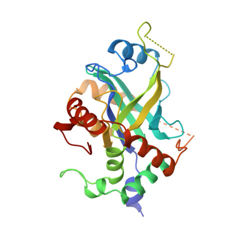
    Human cytomegalovirus (HCMV) protease belongs to a new class of serine proteases, with a unique polypeptide backbone fold. The crystal structure of the protease in complex with a peptidomimetic inhibitor (based on the natural substrates and covering the P4 to P1' positions) has been determined at 2.7 A resolution. The inhibitor is bound in an extended conformation, forming an anti-parallel beta-sheet with the protease. The P3 and P1 side chains are less accessible to solvent, whereas the P4 and P2 side chains are more exposed. The inhibitor binding mode shows significant similarity to those observed for peptidomimetic inhibitors or substrates of other classes of serine proteases (chymotrypsin and subtilisin). HCMV protease therefore represents example of convergent evolution. In addition, large conformational differences relative to the structure of the free enzyme are observed, which may be important for inhibitor binding.


  • Organizational Affiliation

    Boehringer Ingelheim Pharmaceuticals, Inc., Ridgefield, Connecticut 06877, USA. tong@como.bio.columbia.edu


Macromolecules
Find similar proteins by:  (by identity cutoff)  |  3D Structure
Entity ID: 1
MoleculeChains Sequence LengthOrganismDetailsImage
HUMAN CYTOMEGALOVIRUS PROTEASE
A, B, C, D
256Human betaherpesvirus 5Mutation(s): 3 
EC: 3.4.21
UniProt
Find proteins for P16753 (Human cytomegalovirus (strain AD169))
Explore P16753 
Go to UniProtKB:  P16753
Entity Groups  
Sequence Clusters30% Identity50% Identity70% Identity90% Identity95% Identity100% Identity
UniProt GroupP16753
Sequence Annotations
Expand
  • Reference Sequence
Small Molecules
Ligands 1 Unique
IDChains Name / Formula / InChI Key2D Diagram3D Interactions
01E
Query on 01E

Download Ideal Coordinates CCD File 
E [auth A],
F [auth B],
G [auth C],
H [auth D]
(2S)-2-(3,3-dimethylbutanoylamino)-N-[(2S)-1-[[(2S,3S)-3-hydroxy-4-[(4-iodophenyl)methylamino]-4-oxo-butan-2-yl]amino]- 1,4-dioxo-4-pyrrol-1-yl-butan-2-yl]-3,3-dimethyl-butanamide
C31 H44 I N5 O6
DUBMDYQWHSIBPD-FBESYSLLSA-N
Biologically Interesting Molecules (External Reference) 1 Unique
Entity ID: 2
IDChains NameType/Class2D Diagram3D Interactions
PRD_000229 (01E)
Query on PRD_000229
E [auth A],
F [auth B],
G [auth C],
H [auth D]
BILC 821Peptide-like / Inhibitor
Experimental Data & Validation

Experimental Data

  • Method: X-RAY DIFFRACTION
  • Resolution: 2.70 Å
  • R-Value Free: 0.332 
  • R-Value Work: 0.226 
  • R-Value Observed: 0.226 
  • Space Group: P 21 2 21
Unit Cell:
Length ( Å )Angle ( ˚ )
a = 107.8α = 90
b = 53.4β = 90
c = 212.4γ = 90
Software Package:
Software NamePurpose
REPLACEmodel building
X-PLORrefinement
DENZOdata reduction
SCALEPACKdata scaling
REPLACEphasing

Structure Validation

View Full Validation Report


Entry History 

Deposition Data

Revision History  (Full details and data files)

  • Version 1.0: 1999-08-04
    Type: Initial release
  • Version 1.1: 2008-03-25
    Changes: Version format compliance
  • Version 1.2: 2011-07-13
    Changes: Atomic model, Database references, Derived calculations, Non-polymer description, Source and taxonomy, Structure summary, Version format compliance
  • Version 1.3: 2012-12-12
    Changes: Other
  • Version 1.4: 2021-11-03
    Changes: Database references, Derived calculations, Other, Structure summary
  • Version 1.5: 2023-08-09
    Changes: Refinement description