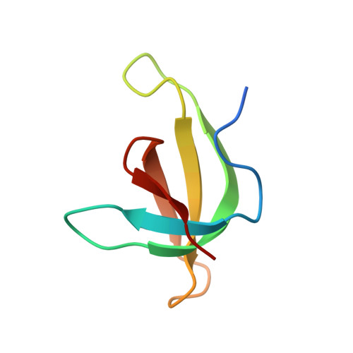
Structure of the Q67H mutant of R67 dihydrofolate reductase-NADP+ complex reveals a novel cofactor binding mode.
Divya, N., Grifith, E., Narayana, N.(2007) Protein Sci 16: 1063-1068
- PubMed: 17473013
- DOI: https://doi.org/10.1110/ps.062740907
- Primary Citation of Related Structures:
2P4T - PubMed Abstract:
Plasmid-encoded bacterial R67 dihydrofolate reductase (DHFR) is a NADPH-dependent enzyme unrelated to chromosomal DHFR in amino acid sequence and structure. R67 DHFR is insensitive to the bacterial drug trimethoprim in contrast to chromosomal DHFR. The crystal structure of Q67H mutant of R67 DHFR bound to NADP(+) has been determined at 1.15 angstroms resolution. The cofactor assumes an extended conformation with the nicotinamide ring bound near the center of the active site pore, the ribose and pyrophosphate group (PP(i)) extending toward the outer pore. The ribonicotinamide exhibits anti conformation as in chromosomal DHFR complexes. The relative orientation between the PP(i) and the nicotinamide ribose differs from that observed in chromosomal DHFR-NADP(+) complexes. The coenzyme displays symmetrical binding mode with several water-mediated hydrogen bonds with the protein besides ionic, stacking, and van der Waals interactions. The structure provides a molecular basis for the observed stoichiometry and cooperativity in ligand binding. The ternary model based on the present structure and the previous R67 DHFR-folate complex provides insight into the catalytic mechanism and indicates that the relative orientation of the reactants in plasmid DHFR is different from that seen in chromosomal DHFRs.
Organizational Affiliation:
Department of Biochemistry, Case Western Reserve University, Cleveland, Ohio 44106, USA.