Spatial Structure and pH-dependent Conformational Diversity of Dimeric Transmembrane Domain of the Receptor Tyrosine Kinase EphA1
Bocharov, E.V., Mayzel, M.L., Volynsky, P.E., Goncharuk, M.V., Ermolyuk, Y.S., Schulga, A.A., Artemenko, E.O., Efremov, R.G., Arseniev, A.S.(2008) J Biol Chem 283: 29385-29395
- PubMed: 18728013
- DOI: https://doi.org/10.1074/jbc.M803089200
- Primary Citation of Related Structures:
2K1K, 2K1L - PubMed Abstract:
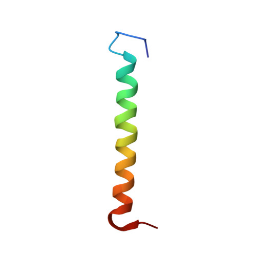
Eph receptors are found in a wide variety of cells in developing and mature tissues and represent the largest family of receptor tyrosine kinases, regulating cell shape, movements, and attachment. The receptor tyrosine kinases conduct biochemical signals across plasma membrane via lateral dimerization in which their transmembrane domains play an important role. Structural-dynamic properties of the homodimeric transmembrane domain of the EphA1 receptor were investigated with the aid of solution NMR in lipid bicelles and molecular dynamics in explicit lipid bilayer. EphA1 transmembrane segments associate in a right-handed parallel alpha-helical bundle, region (544-569)(2), through the N-terminal glycine zipper motif A(550)X(3)G(554)X(3)G(558). Under acidic conditions, the N terminus of the transmembrane helix is stabilized by an N-capping box formed by the uncharged carboxyl group of Glu(547), whereas its deprotonation results in a rearrangement of hydrogen bonds, fractional unfolding of the helix, and a realignment of the helix-helix packing with appearance of additional minor dimer conformation utilizing seemingly the C-terminal GG4-like dimerization motif A(560)X(3)G(564). This can be interpreted as the ability of the EphA1 receptor to adjust its response to ligand binding according to extracellular pH. The dependence of the pK(a) value of Glu(547) and the dimer conformational equilibrium on the lipid head charge suggests that both local environment and membrane surface potential can modulate dimerization and activation of the receptor. This makes the EphA1 receptor unique among the Eph family, implying its possible physiological role as an "extracellular pH sensor," and can have relevant physiological implications.
Organizational Affiliation:
Division of Structural Biology, Shemyakin-Ovchinnikov Institute of Bioorganic Chemistry, Russian Academy of Sciences, Ul. Miklukho-Maklaya, 16/10, Moscow 117997, Russia. bon@nmr.ru