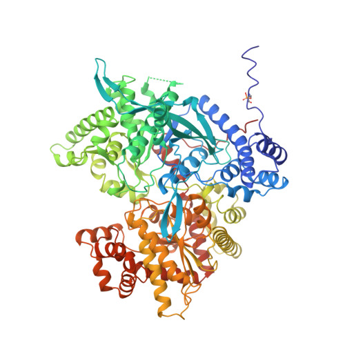
Allosteric inhibition of glycogen phosphorylase a by the potential antidiabetic drug 3-isopropyl 4-(2-chlorophenyl)-1,4-dihydro-1-ethyl-2-methyl-pyridine-3,5,6-tricarbo xylate.
Oikonomakos, N.G., Tsitsanou, K.E., Zographos, S.E., Skamnaki, V.T., Goldmann, S., Bischoff, H.(1999) Protein Sci 8: 1930-1945
- PubMed: 10548038
- DOI: https://doi.org/10.1110/ps.8.10.1930
- Primary Citation of Related Structures:
2GPA, 3AMV - PubMed Abstract:
The effect of the potential antidiabetic drug (-)(S)-3-isopropyl 4-(2-chlorophenyl)-1,4-dihydro-1-ethyl-2-methyl-pyridine-3,5,6-tricarbox ylate (W1807) on the catalytic and structural properties of glycogen phosphorylase a has been studied. Glycogen phosphorylase (GP) is an allosteric enzyme whose activity is primarily controlled by reversible phosphorylation of Ser14 of the dephosphorylated enzyme (GPb, less active, predominantly T-state) to form the phosphorylated enzyme (GPa, more active, predominantly R-state). Upon conversion of GPb to GPa, the N-terminal tail (residues 5-22), which carries the Ser14(P), changes its conformation into a distorted 3(10) helix and its contacts from intrasubunit to intersubunit. This alteration causes a series of tertiary and quaternary conformational changes that lead to activation of the enzyme through opening access to the catalytic site. As part of a screening process to identify compounds that might contribute to the regulation of glycogen metabolism in the noninsulin dependent diabetes diseased state, W1807 has been found as the most potent inhibitor of GPb (Ki = 1.6 nM) that binds at the allosteric site of T-state GPb and produces further conformational changes, characteristic of a T'-like state. Kinetics show W1807 is a potent competitive inhibitor of GPa (-AMP) (Ki = 10.8 nM) and of GPa (+1 mM AMP) (Ki = 19.4 microM) with respect to glucose 1-phosphate and acts in synergism with glucose. To elucidate the structural features that contribute to the binding, the structures of GPa in the T-state conformation in complex with glucose and in complex with both glucose and W1807 have been determined at 100 K to 2.0 A and 2.1 A resolution, and refined to crystallographic R-values of 0.179 (R(free) = 0.230) and 0.189 (R(free) = 0.263), respectively. W1807 binds tightly at the allosteric site and induces substantial conformational changes both in the vicinity of the allosteric site and the subunit interface. A disordering of the N-terminal tail occurs, while the loop of chain containing residues 192-196 and residues 43'-49' shift to accommodate the ligand. Structural comparisons show that the T-state GPa-glucose-W1807 structure is overall more similar to the T-state GPb-W1807 complex structure than to the GPa-glucose complex structure, indicating that W1807 is able to transform GPa to the T'-like state already observed with GPb. The structures provide a rational for the potency of the inhibitor and explain GPa allosteric inhibition of activity upon W1807 binding.
Organizational Affiliation:
Institute of Biological Research and Biotechnology, The National Hellenic Research, Athens, Greece. nikos@aegean.eie.gr