2ZB5

Crystal structure of the measles virus hemagglutinin (complex-sugar-type)


Experimental Data Snapshot

  • Method: X-RAY DIFFRACTION
  • Resolution: 3.00 Å
  • R-Value Free: 0.271 
  • R-Value Work: 0.227 

wwPDB Validation   3D Report Full Report


Ligand Structure Quality Assessment 


This is version 1.3 of the entry. See complete history


Literature

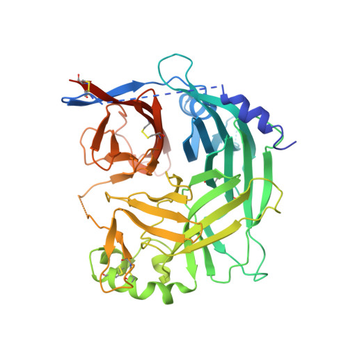
Crystal structure of measles virus hemagglutinin provides insight into effective vaccines

Hashiguchi, T.Kajikawa, M.Maita, N.Takeda, M.Kuroki, K.Sasaki, K.Kohda, D.Yanagi, Y.Maenaka, K.

(2007) Proc Natl Acad Sci U S A 104: 19535-19540

  • DOI: https://doi.org/10.1073/pnas.0707830104
  • Primary Citation of Related Structures:  
    2ZB5, 2ZB6

  • PubMed Abstract: 

    Measles still remains a major cause of childhood morbidity and mortality worldwide. Measles virus (MV) vaccines are highly successful, but the mechanism underlying their efficacy has been unclear. Here we report the crystal structure of the MV attachment protein, hemagglutinin, responsible for MV entry. The receptor-binding head domain exhibits a cubic-shaped beta-propeller structure and forms a homodimer. N-linked sugars appear to mask the broad regions and cause the two molecules forming the dimer to tilt oppositely toward the horizontal plane. Accordingly, residues of the putative receptor-binding site, highly conserved among MV strains, are strategically positioned in the unshielded area of the protein. These conserved residues also serve as epitopes for neutralizing antibodies, ensuring the serological monotype, a basis for effective MV vaccines. Our findings suggest that sugar moieties in the MV hemagglutinin critically modulate virus-receptor interaction as well as antiviral antibody responses, differently from sugars of the HIV gp120, which allow for immune evasion.


  • Organizational Affiliation

    Department of Virology, Faculty of Medicine, and Division of Structural Biology, Medical Institute of Bioregulation, Kyushu University, 3-1-1 Maidashi, Higashi-ku, Fukuoka, Fukuoka 812-8582, Japan.


Macromolecules
Find similar proteins by:  (by identity cutoff)  |  3D Structure
Entity ID: 1
MoleculeChains Sequence LengthOrganismDetailsImage
Hemagglutinin protein481Measles virus strain Edmonston-BMutation(s): 2 
UniProt
Find proteins for P08362 (Measles virus (strain Edmonston))
Explore P08362 
Go to UniProtKB:  P08362
Entity Groups  
Sequence Clusters30% Identity50% Identity70% Identity90% Identity95% Identity100% Identity
UniProt GroupP08362
Sequence Annotations
Expand
  • Reference Sequence
Small Molecules
Ligands 1 Unique
IDChains Name / Formula / InChI Key2D Diagram3D Interactions
NAG
Query on NAG

Download Ideal Coordinates CCD File 
B [auth A],
C [auth A]
2-acetamido-2-deoxy-beta-D-glucopyranose
C8 H15 N O6
OVRNDRQMDRJTHS-FMDGEEDCSA-N
Experimental Data & Validation

Experimental Data

  • Method: X-RAY DIFFRACTION
  • Resolution: 3.00 Å
  • R-Value Free: 0.271 
  • R-Value Work: 0.227 
  • Space Group: P 41 21 2
Unit Cell:
Length ( Å )Angle ( ˚ )
a = 134.523α = 90
b = 134.523β = 90
c = 99.251γ = 90
Software Package:
Software NamePurpose
HKL-2000data collection
SOLVEphasing
CNSrefinement
HKL-2000data reduction
HKL-2000data scaling

Structure Validation

View Full Validation Report


Ligand Structure Quality Assessment 


Entry History 

Deposition Data

Revision History  (Full details and data files)

  • Version 1.0: 2007-11-06
    Type: Initial release
  • Version 1.1: 2011-07-13
    Changes: Non-polymer description, Version format compliance
  • Version 1.2: 2020-07-29
    Type: Remediation
    Reason: Carbohydrate remediation
    Changes: Data collection, Database references, Derived calculations, Structure summary
  • Version 1.3: 2021-11-10
    Changes: Database references, Structure summary