2YOC

Crystal structure of PulA from Klebsiella oxytoca


Experimental Data Snapshot

  • Method: X-RAY DIFFRACTION
  • Resolution: 2.88 Å
  • R-Value Free: 0.213 
  • R-Value Work: 0.171 
  • R-Value Observed: 0.173 

wwPDB Validation   3D Report Full Report


This is version 1.3 of the entry. See complete history


Literature

Structural Basis of Pullulanase Membrane Binding and Secretion Revealed by X-Ray Crystallography, Molecular Dynamics and Biochemical Analysis

East, A.Mechaly, A.E.Huysmans, G.H.M.Bernarde, C.Tello-Manigne, D.Nadeau, N.Pugsley, A.P.Buschiazzo, A.Alzari, P.M.Bond, P.J.Francetic, O.

(2016) Structure 24: 92

  • DOI: https://doi.org/10.1016/j.str.2015.10.023
  • Primary Citation of Related Structures:  
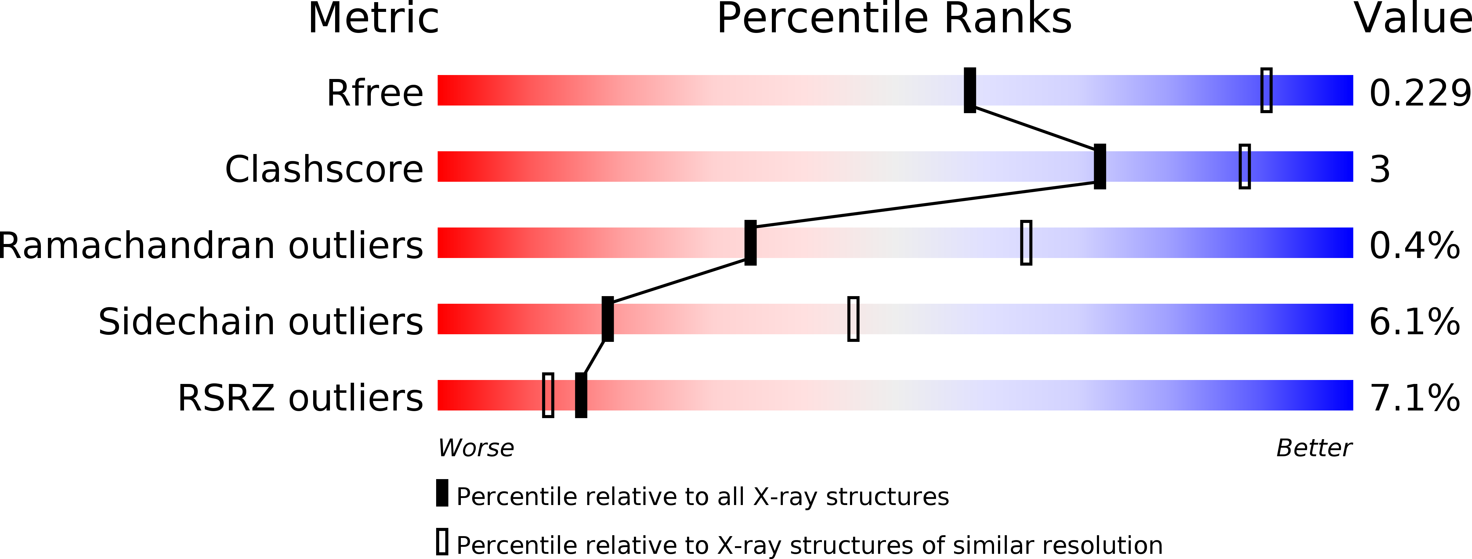
    2YOC

  • PubMed Abstract: 

    The Klebsiella lipoprotein pullulanase (PulA) is exported to the periplasm, triacylated, and anchored via lipids in the inner membrane (IM) prior to its transport to the bacterial surface through a type II secretion system (T2SS). X-Ray crystallography and atomistic molecular dynamics (MD) simulations of PulA in a 1-palmitoyl-2-oleoyl-sn-glycero-3-phosphoethanolamine (POPE) model membrane provided an unprecedented molecular view of an N-terminal unstructured tether and the IM lipoprotein retention signal, and revealed novel interactions with the IM via N-terminal immunoglobulin-like domains in PulA. An efficiently secreted nonacylated variant (PulANA) showed similar peripheral membrane association during MD simulations, consistent with the binding of purified PulANA to liposomes. Remarkably, combined X-ray, MD, and functional studies identified a novel subdomain, Ins, inserted in the α-amylase domain, which is required for PulA secretion. Available data support a model in which PulA binding to the IM promotes interactions with the T2SS, possibly via the Ins subdomain.


  • Organizational Affiliation

    Department of Chemistry, Centre for Molecular Informatics, University of Cambridge, Lensfield Road, Cambridge CB2 1EW, UK.


Macromolecules
Find similar proteins by:  (by identity cutoff)  |  3D Structure
Entity ID: 1
MoleculeChains Sequence LengthOrganismDetailsImage
PULLULANASE
A, B
1,078Klebsiella oxytocaMutation(s): 0 
EC: 3.2.1.41
Membrane Entity: Yes 
UniProt
Find proteins for P07206 (Klebsiella pneumoniae)
Explore P07206 
Go to UniProtKB:  P07206
Entity Groups  
Sequence Clusters30% Identity50% Identity70% Identity90% Identity95% Identity100% Identity
UniProt GroupP07206
Sequence Annotations
Expand
  • Reference Sequence
Small Molecules
Ligands 2 Unique
IDChains Name / Formula / InChI Key2D Diagram3D Interactions
SO4
Query on SO4

Download Ideal Coordinates CCD File 
G [auth A]
H [auth A]
I [auth A]
J [auth A]
O [auth B]
G [auth A],
H [auth A],
I [auth A],
J [auth A],
O [auth B],
P [auth B],
Q [auth B],
R [auth B],
S [auth B],
T [auth B],
U [auth B],
V [auth B],
W [auth B],
X [auth B]
SULFATE ION
O4 S
QAOWNCQODCNURD-UHFFFAOYSA-L
CA
Query on CA

Download Ideal Coordinates CCD File 
C [auth A]
D [auth A]
E [auth A]
F [auth A]
K [auth B]
C [auth A],
D [auth A],
E [auth A],
F [auth A],
K [auth B],
L [auth B],
M [auth B],
N [auth B]
CALCIUM ION
Ca
BHPQYMZQTOCNFJ-UHFFFAOYSA-N
Modified Residues  1 Unique
IDChains TypeFormula2D DiagramParent
MSE
Query on MSE
A, B
L-PEPTIDE LINKINGC5 H11 N O2 SeMET
Experimental Data & Validation

Experimental Data

  • Method: X-RAY DIFFRACTION
  • Resolution: 2.88 Å
  • R-Value Free: 0.213 
  • R-Value Work: 0.171 
  • R-Value Observed: 0.173 
  • Space Group: P 63 2 2
Unit Cell:
Length ( Å )Angle ( ˚ )
a = 179.15α = 90
b = 179.15β = 90
c = 334.69γ = 120
Software Package:
Software NamePurpose
BUSTERrefinement
XDSdata reduction
SCALAdata scaling

Structure Validation

View Full Validation Report


Entry History 

Deposition Data

Revision History  (Full details and data files)

  • Version 1.0: 2013-11-06
    Type: Initial release
  • Version 1.1: 2015-12-23
    Changes: Database references
  • Version 1.2: 2016-01-13
    Changes: Database references
  • Version 1.3: 2016-01-20
    Changes: Database references