2X2E

Dynamin GTPase dimer, long axis form


Experimental Data Snapshot

  • Method: X-RAY DIFFRACTION
  • Resolution: 2.00 Å
  • R-Value Free: 0.230 
  • R-Value Work: 0.185 
  • R-Value Observed: 0.185 

wwPDB Validation   3D Report Full Report


Ligand Structure Quality Assessment 


This is version 1.2 of the entry. See complete history


Literature

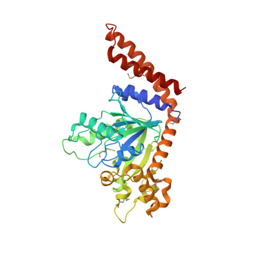
G Domain Dimerization Controls Dynamin'S Assembly-Stimulated Gtpase Activity.

Chappie, J.S.Acharya, S.Leonard, M.Schmid, S.L.Dyda, F.

(2010) Nature 465: 435

  • DOI: https://doi.org/10.1038/nature09032
  • Primary Citation of Related Structures:  
    2X2E, 2X2F

  • PubMed Abstract: 

    Dynamin is an atypical GTPase that catalyses membrane fission during clathrin-mediated endocytosis. The mechanisms of dynamin's basal and assembly-stimulated GTP hydrolysis are unknown, though both are indirectly influenced by the GTPase effector domain (GED). Here we present the 2.0 A resolution crystal structure of a human dynamin 1-derived minimal GTPase-GED fusion protein, which was dimeric in the presence of the transition state mimic GDP.AlF(4)(-).The structure reveals dynamin's catalytic machinery and explains how assembly-stimulated GTP hydrolysis is achieved through G domain dimerization. A sodium ion present in the active site suggests that dynamin uses a cation to compensate for the developing negative charge in the transition state in the absence of an arginine finger. Structural comparison to the rat dynamin G domain reveals key conformational changes that promote G domain dimerization and stimulated hydrolysis. The structure of the GTPase-GED fusion protein dimer provides insight into the mechanisms underlying dynamin-catalysed membrane fission.


  • Organizational Affiliation

    Laboratory of Molecular Biology, National Institute of Diabetes and Digestive and Kidney Diseases, NIH, Bethesda, Maryland 20892, USA.


Macromolecules
Find similar proteins by:  (by identity cutoff)  |  3D Structure
Entity ID: 1
MoleculeChains Sequence LengthOrganismDetailsImage
DYNAMIN-1A,
B [auth D]
353Homo sapiensMutation(s): 1 
EC: 3.6.5.5
UniProt & NIH Common Fund Data Resources
Find proteins for Q05193 (Homo sapiens)
Explore Q05193 
Go to UniProtKB:  Q05193
PHAROS:  Q05193
GTEx:  ENSG00000106976 
Entity Groups  
Sequence Clusters30% Identity50% Identity70% Identity90% Identity95% Identity100% Identity
UniProt GroupQ05193
Sequence Annotations
Expand
  • Reference Sequence
Small Molecules
Ligands 4 Unique
IDChains Name / Formula / InChI Key2D Diagram3D Interactions
GDP
Query on GDP

Download Ideal Coordinates CCD File 
C [auth A],
G [auth D]
GUANOSINE-5'-DIPHOSPHATE
C10 H15 N5 O11 P2
QGWNDRXFNXRZMB-UUOKFMHZSA-N
ALF
Query on ALF

Download Ideal Coordinates CCD File 
D [auth A],
H [auth D]
TETRAFLUOROALUMINATE ION
Al F4
UYOMQIYKOOHAMK-UHFFFAOYSA-J
MG
Query on MG

Download Ideal Coordinates CCD File 
E [auth A],
I [auth D]
MAGNESIUM ION
Mg
JLVVSXFLKOJNIY-UHFFFAOYSA-N
NA
Query on NA

Download Ideal Coordinates CCD File 
F [auth A],
J [auth D]
SODIUM ION
Na
FKNQFGJONOIPTF-UHFFFAOYSA-N
Modified Residues  1 Unique
IDChains TypeFormula2D DiagramParent
MSE
Query on MSE
A,
B [auth D]
L-PEPTIDE LINKINGC5 H11 N O2 SeMET
Experimental Data & Validation

Experimental Data

  • Method: X-RAY DIFFRACTION
  • Resolution: 2.00 Å
  • R-Value Free: 0.230 
  • R-Value Work: 0.185 
  • R-Value Observed: 0.185 
  • Space Group: P 21 21 21
Unit Cell:
Length ( Å )Angle ( ˚ )
a = 43.61α = 90
b = 81.02β = 90
c = 181.54γ = 90
Software Package:
Software NamePurpose
XDSdata reduction
XSCALEdata scaling
SHARPDphasing
DMphasing
CNSrefinement

Structure Validation

View Full Validation Report


Ligand Structure Quality Assessment 


Entry History 

Deposition Data

Revision History  (Full details and data files)

  • Version 1.0: 2010-04-28
    Type: Initial release
  • Version 1.1: 2011-08-31
    Changes: Database references, Other, Refinement description, Structure summary, Version format compliance
  • Version 1.2: 2019-05-08
    Changes: Data collection, Derived calculations, Experimental preparation, Other