2VER

Structural model for the complex between the Dr adhesins and carcinoembryonic antigen (CEA)


Experimental Data Snapshot

  • Method: SOLUTION NMR
  • Conformers Calculated: 200 
  • Conformers Submitted: 
  • Selection Criteria: REPRESENTATIVE STRUCTURE 

wwPDB Validation   3D Report Full Report


This is version 1.2 of the entry. See complete history


Literature

Binding of Dr Adhesins of Escherichia Coli to Carcinoembryonic Antigen Triggers Receptor Dissociation.

Korotkova, N.Yang, Y.Le Trong, I.Cota, E.Demeler, B.Marchant, J.Thomas, W.E.Stenkamp, R.E.Moseley, S.L.Matthews, S.

(2008) Mol Microbiol 67: 420

  • DOI: https://doi.org/10.1111/j.1365-2958.2007.06054.x
  • Primary Citation of Related Structures:  
    2QSQ, 2QST, 2VER

  • PubMed Abstract: 

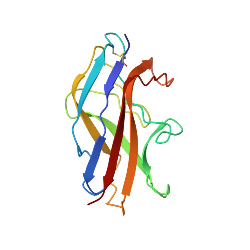
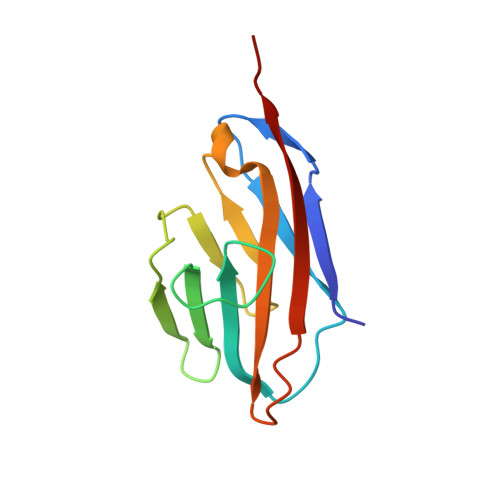
    Carcinoembryonic antigen (CEA)-related cell adhesion molecules (CEACAMs) are host receptors for the Dr family of adhesins of Escherichia coli. To define the mechanism for binding of Dr adhesins to CEACAM receptors, we carried out structural studies on the N-terminal domain of CEA and its complex with the Dr adhesin. The crystal structure of CEA reveals a dimer similar to other dimers formed by receptors with IgV-like domains. The structure of the CEA/Dr adhesin complex is proposed based on NMR spectroscopy and mutagenesis data in combination with biochemical characterization. The Dr adhesin/CEA interface overlaps appreciably with the region responsible for CEA dimerization. Binding kinetics, mutational analysis and spectroscopic examination of CEA dimers suggest that Dr adhesins can dissociate CEA dimers prior to the binding of monomeric forms. Our conclusions include a plausible mechanism for how E. coli, and perhaps other bacterial and viral pathogens, exploit CEACAMs. The present structure of the complex provides a powerful tool for the design of novel inhibitory strategies to treat E. coli infections.


  • Organizational Affiliation

    Department of Microbiology, University of Washington, Seattle, WA 98195-7242, USA.


Macromolecules
Find similar proteins by:  (by identity cutoff)  |  3D Structure
Entity ID: 1
MoleculeChains Sequence LengthOrganismDetailsImage
AFIMBRIAL ADHESIN AFA-III143Escherichia coliMutation(s): 0 
UniProt
Find proteins for Q57254 (Escherichia coli)
Explore Q57254 
Go to UniProtKB:  Q57254
Entity Groups  
Sequence Clusters30% Identity50% Identity70% Identity90% Identity95% Identity100% Identity
UniProt GroupQ57254
Sequence Annotations
Expand
  • Reference Sequence
Find similar proteins by:  (by identity cutoff)  |  3D Structure
Entity ID: 2
MoleculeChains Sequence LengthOrganismDetailsImage
ARCINOEMBRYONIC ANTIGEN-RELATED CELL ADHESION MOLECULE 5B [auth N]110Homo sapiensMutation(s): 0 
UniProt & NIH Common Fund Data Resources
Find proteins for P06731 (Homo sapiens)
Explore P06731 
Go to UniProtKB:  P06731
PHAROS:  P06731
GTEx:  ENSG00000105388 
Entity Groups  
Sequence Clusters30% Identity50% Identity70% Identity90% Identity95% Identity100% Identity
UniProt GroupP06731
Sequence Annotations
Expand
  • Reference Sequence
Small Molecules
Ligands 1 Unique
IDChains Name / Formula / InChI Key2D Diagram3D Interactions
MTN
Query on MTN

Download Ideal Coordinates CCD File 
C [auth N],
D [auth N],
E [auth N]
S-[(1-oxyl-2,2,5,5-tetramethyl-2,5-dihydro-1H-pyrrol-3-yl)methyl] methanesulfonothioate
C10 H18 N O3 S2
MXZPGYFBZHBAQM-UHFFFAOYSA-N
Experimental Data & Validation

Experimental Data

  • Method: SOLUTION NMR
  • Conformers Calculated: 200 
  • Conformers Submitted: 
  • Selection Criteria: REPRESENTATIVE STRUCTURE 

Structure Validation

View Full Validation Report


Entry History 

Deposition Data

Revision History  (Full details and data files)

  • Version 1.0: 2008-01-08
    Type: Initial release
  • Version 1.1: 2011-05-12
    Changes: Version format compliance
  • Version 1.2: 2011-07-13
    Changes: Version format compliance