2VED

crystal structure of the chimerical mutant CapABK55M protein


Experimental Data Snapshot

  • Method: X-RAY DIFFRACTION
  • Resolution: 2.60 Å
  • R-Value Free: 0.270 
  • R-Value Work: 0.204 
  • R-Value Observed: 0.208 

wwPDB Validation   3D Report Full Report


Ligand Structure Quality Assessment 


This is version 1.3 of the entry. See complete history


Literature

Structural Basis for the Regulation Mechanism of the Tyrosine Kinase Capb from Staphylococcus Aureus.

Olivares-Illana, V.Meyer, P.Bechet, E.Gueguen-Chaignon, V.Soulat, D.Lazereg-Riquier, S.Mijakovic, I.Deutscher, J.Cozzone, A.J.Laprevote, O.Morera, S.Grangeasse, C.Nessler, S.

(2008) PLoS Biol 6: E143

  • DOI: https://doi.org/10.1371/journal.pbio.0060143
  • Primary Citation of Related Structures:  
    2VED, 3BFV

  • PubMed Abstract: 

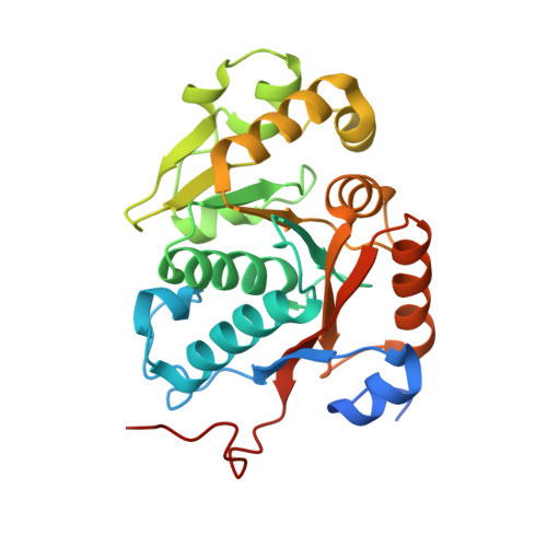
    Bacteria were thought to be devoid of tyrosine-phosphorylating enzymes. However, several tyrosine kinases without similarity to their eukaryotic counterparts have recently been identified in bacteria. They are involved in many physiological processes, but their accurate functions remain poorly understood due to slow progress in their structural characterization. They have been best characterized as copolymerases involved in the synthesis and export of extracellular polysaccharides. These compounds play critical roles in the virulence of pathogenic bacteria, and bacterial tyrosine kinases can thus be considered as potential therapeutic targets. Here, we present the crystal structures of the phosphorylated and unphosphorylated states of the tyrosine kinase CapB from the human pathogen Staphylococcus aureus together with the activator domain of its cognate transmembrane modulator CapA. This first high-resolution structure of a bacterial tyrosine kinase reveals a 230-kDa ring-shaped octamer that dissociates upon intermolecular autophosphorylation. These observations provide a molecular basis for the regulation mechanism of the bacterial tyrosine kinases and give insights into their copolymerase function.


  • Organizational Affiliation

    Laboratoire d'Enzymologie et Biochimie Structurales, CNRS, Gif sur Yvette, France.


Macromolecules
Find similar proteins by:  (by identity cutoff)  |  3D Structure
Entity ID: 1
MoleculeChains Sequence LengthOrganismDetailsImage
MEMBRANE PROTEIN CAPA1, PROTEIN TYROSINE KINASE
A, B
271Staphylococcus aureusMutation(s): 1 
Gene Names: CAPA1CAPB2
UniProt
Find proteins for A8YPQ6 (Staphylococcus aureus)
Explore A8YPQ6 
Go to UniProtKB:  A8YPQ6
Find proteins for A8YPQ5 (Staphylococcus aureus)
Explore A8YPQ5 
Go to UniProtKB:  A8YPQ5
Entity Groups  
Sequence Clusters30% Identity50% Identity70% Identity90% Identity95% Identity100% Identity
UniProt GroupsA8YPQ6A8YPQ5
Sequence Annotations
Expand
  • Reference Sequence
Experimental Data & Validation

Experimental Data

  • Method: X-RAY DIFFRACTION
  • Resolution: 2.60 Å
  • R-Value Free: 0.270 
  • R-Value Work: 0.204 
  • R-Value Observed: 0.208 
  • Space Group: I 4
Unit Cell:
Length ( Å )Angle ( ˚ )
a = 163.2α = 90
b = 163.2β = 90
c = 57.2γ = 90
Software Package:
Software NamePurpose
REFMACrefinement
MOSFLMdata reduction
SCALAdata scaling
PHASERphasing

Structure Validation

View Full Validation Report


Ligand Structure Quality Assessment 


Entry History 

Deposition Data

Revision History  (Full details and data files)

  • Version 1.0: 2008-07-01
    Type: Initial release
  • Version 1.1: 2011-05-08
    Changes: Version format compliance
  • Version 1.2: 2011-07-13
    Changes: Version format compliance
  • Version 1.3: 2023-12-13
    Changes: Data collection, Database references, Derived calculations, Other, Refinement description