2REA

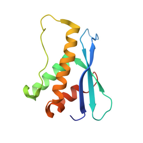
Crystal structures of C2ALPHA-PI3 kinase PX-domain domain indicate conformational change associated with ligand binding.


Experimental Data Snapshot

  • Method: X-RAY DIFFRACTION
  • Resolution: 2.50 Å
  • R-Value Free: 0.318 
  • R-Value Work: 0.231 
  • R-Value Observed: 0.235 

wwPDB Validation   3D Report Full Report


This is version 1.2 of the entry. See complete history


Literature

Crystal structures of PI3K-C2alpha PX domain indicate conformational change associated with ligand binding

Parkinson, G.N.Vines, D.Driscoll, P.C.Djordjevic, S.

(2008) BMC Struct Biol 8: 13-13

  • DOI: https://doi.org/10.1186/1472-6807-8-13
  • Primary Citation of Related Structures:  
    2REA, 2RED

  • PubMed Abstract: 

    PX domains have specialized protein structures involved in binding of phosphoinositides (PIs). Through binding to the various PIs PX domains provide site-specific membrane signals to modulate the intracellular localisation and biological activity of effector proteins. Several crystal structures of these domains are now available from a variety of proteins. All PX domains contain a canonical core structure with main differences exhibited within the loop regions forming the phosphoinositide binding pockets. It is within these areas that the molecular basis for ligand specificity originates.


  • Organizational Affiliation

    Cancer Research UK Biomolecular Structure Group, The School of Pharmacy, University of London, 29-39 Brunswick Square, London WC1N 1AX, UK. gary.parkinson@pharmacy.ac.uk


Macromolecules
Find similar proteins by:  (by identity cutoff)  |  3D Structure
Entity ID: 1
MoleculeChains Sequence LengthOrganismDetailsImage
Phosphatidylinositol-4-phosphate 3-kinase C2 domain-containing alpha polypeptide121Homo sapiensMutation(s): 0 
Gene Names: PIK3C2A
EC: 2.7.1.154
UniProt & NIH Common Fund Data Resources
Find proteins for O00443 (Homo sapiens)
Explore O00443 
Go to UniProtKB:  O00443
PHAROS:  O00443
GTEx:  ENSG00000011405 
Entity Groups  
Sequence Clusters30% Identity50% Identity70% Identity90% Identity95% Identity100% Identity
UniProt GroupO00443
Sequence Annotations
Expand
  • Reference Sequence
Experimental Data & Validation

Experimental Data

  • Method: X-RAY DIFFRACTION
  • Resolution: 2.50 Å
  • R-Value Free: 0.318 
  • R-Value Work: 0.231 
  • R-Value Observed: 0.235 
  • Space Group: P 31 2 1
Unit Cell:
Length ( Å )Angle ( ˚ )
a = 56.866α = 90
b = 56.866β = 90
c = 92.995γ = 120
Software Package:
Software NamePurpose
EPMRphasing
REFMACrefinement
DENZOdata reduction
SCALEPACKdata scaling

Structure Validation

View Full Validation Report


Entry History 

Deposition Data

Revision History  (Full details and data files)

  • Version 1.0: 2007-11-27
    Type: Initial release
  • Version 1.1: 2011-07-13
    Changes: Version format compliance
  • Version 1.2: 2023-08-30
    Changes: Data collection, Database references, Refinement description