2R7G

Structure of the retinoblastoma protein pocket domain in complex with adenovirus E1A CR1 domain


Experimental Data Snapshot

  • Method: X-RAY DIFFRACTION
  • Resolution: 1.67 Å
  • R-Value Free: 0.218 
  • R-Value Work: 0.195 
  • R-Value Observed: 0.198 

wwPDB Validation   3D Report Full Report


This is version 1.4 of the entry. See complete history


Literature

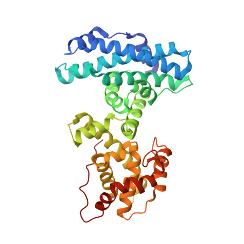
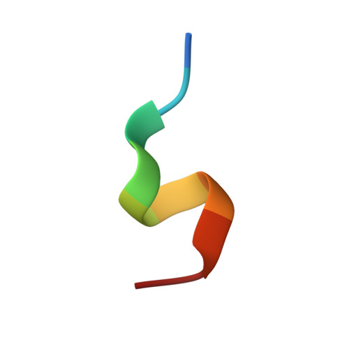
Structure of the retinoblastoma protein bound to adenovirus E1A reveals the molecular basis for viral oncoprotein inactivation of a tumor suppressor

Liu, X.Marmorstein, R.

(2007) Genes Dev 21: 2711-2716

  • DOI: https://doi.org/10.1101/gad.1590607
  • Primary Citation of Related Structures:  
    2R7G

  • PubMed Abstract: 

    The adenovirus (Ad) E1A (Ad-E1A) oncoprotein mediates cell transformation, in part, by displacing E2F transcription factors from the retinoblastoma protein (pRb) tumor suppressor. In this study we determined the crystal structure of the pRb pocket domain in complex with conserved region 1 (CR1) of Ad5-E1A. The structure and accompanying biochemical studies reveal that E1A-CR1 binds at the interface of the A and B cyclin folds of the pRb pocket domain, and that both E1A-CR1 and the E2F transactivation domain use similar conserved nonpolar residues to engage overlapping sites on pRb, implicating a novel molecular mechanism for pRb inactivation by a viral oncoprotein.


  • Organizational Affiliation

    Program in Gene Expression and Regulation, The Wistar Institute, Philadelphia, Pennsylvania 19104, USA.


Macromolecules
Find similar proteins by:  (by identity cutoff)  |  3D Structure
Entity ID: 1
MoleculeChains Sequence LengthOrganismDetailsImage
Retinoblastoma-associated protein
A, C
347Homo sapiensMutation(s): 0 
Gene Names: RB1
UniProt & NIH Common Fund Data Resources
Find proteins for P06400 (Homo sapiens)
Explore P06400 
Go to UniProtKB:  P06400
PHAROS:  P06400
GTEx:  ENSG00000139687 
Entity Groups  
Sequence Clusters30% Identity50% Identity70% Identity90% Identity95% Identity100% Identity
UniProt GroupP06400
Sequence Annotations
Expand
  • Reference Sequence

Find similar proteins by:  Sequence   |   3D Structure  

Entity ID: 2
MoleculeChains Sequence LengthOrganismDetailsImage
Early E1A 32 kDa protein
B, D, E
10Human adenovirus 5Mutation(s): 0 
UniProt
Find proteins for P03255 (Human adenovirus C serotype 5)
Explore P03255 
Go to UniProtKB:  P03255
Entity Groups  
Sequence Clusters30% Identity50% Identity70% Identity90% Identity95% Identity100% Identity
UniProt GroupP03255
Sequence Annotations
Expand
  • Reference Sequence
Experimental Data & Validation

Experimental Data

  • Method: X-RAY DIFFRACTION
  • Resolution: 1.67 Å
  • R-Value Free: 0.218 
  • R-Value Work: 0.195 
  • R-Value Observed: 0.198 
  • Space Group: P 1
Unit Cell:
Length ( Å )Angle ( ˚ )
a = 57.713α = 94.34
b = 57.706β = 92.69
c = 92.268γ = 105.79
Software Package:
Software NamePurpose
DENZOdata reduction
SCALEPACKdata scaling
MOLREPphasing
REFMACrefinement
PDB_EXTRACTdata extraction
HKL-2000data collection
HKL-2000data reduction

Structure Validation

View Full Validation Report


Entry History 

Deposition Data

Revision History  (Full details and data files)

  • Version 1.0: 2007-10-02
    Type: Initial release
  • Version 1.1: 2011-07-13
    Changes: Advisory, Version format compliance
  • Version 1.2: 2017-07-26
    Changes: Refinement description, Source and taxonomy
  • Version 1.3: 2017-10-25
    Changes: Refinement description
  • Version 1.4: 2024-02-21
    Changes: Data collection, Database references, Derived calculations