2R2W

Urokinase plasminogen activator B-chain-GPPE complex


Experimental Data Snapshot

  • Method: X-RAY DIFFRACTION
  • Resolution: 2.01 Å
  • R-Value Free: 0.233 
  • R-Value Work: 0.199 

wwPDB Validation   3D Report Full Report


Ligand Structure Quality Assessment 


This is version 1.2 of the entry. See complete history


Literature

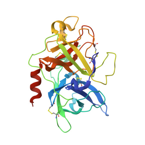
Geometry of GPPE binding to picrate and to the urokinase type plasminogen activator.

Zeslawska, E.Sturzebecher, J.Oleksyn, B.J.

(2007) Bioorg Med Chem Lett 17: 6212-6215

  • DOI: https://doi.org/10.1016/j.bmcl.2007.09.020
  • Primary Citation of Related Structures:  
    2R2W

  • PubMed Abstract: 

    Crystal structure of 2-(4-guanidynephenyl)-1-phenyl-ethanone (GPPE) in two different environments was determined in order to compare the binding geometry of these compound to a simple picrate anion and to protein, urokinase-type plasminogen activator (uPA), which may be treated as a target for anti-cancer drugs. It was shown that the conformation and the hydrogen-bonding formation by GPPE molecule are similar in both environments, but several important differences were discovered and described.


  • Organizational Affiliation

    Department of Chemistry, Pedagogical University, ul. Podchorazych 2, 30-084 Kraków, Poland.


Macromolecules
Find similar proteins by:  (by identity cutoff)  |  3D Structure
Entity ID: 1
MoleculeChains Sequence LengthOrganismDetailsImage
Plasminogen activator, urokinaseA [auth U]253Homo sapiensMutation(s): 1 
Gene Names: PLAU
EC: 3.4.21.73
UniProt & NIH Common Fund Data Resources
Find proteins for P00749 (Homo sapiens)
Explore P00749 
Go to UniProtKB:  P00749
PHAROS:  P00749
GTEx:  ENSG00000122861 
Entity Groups  
Sequence Clusters30% Identity50% Identity70% Identity90% Identity95% Identity100% Identity
UniProt GroupP00749
Sequence Annotations
Expand
  • Reference Sequence
Small Molecules
Ligands 2 Unique
IDChains Name / Formula / InChI Key2D Diagram3D Interactions
4PG
Query on 4PG

Download Ideal Coordinates CCD File 
C [auth U]1-[4-(2-oxo-2-phenylethyl)phenyl]guanidine
C15 H15 N3 O
MERNPSIIBFTCAI-UHFFFAOYSA-N
SO4
Query on SO4

Download Ideal Coordinates CCD File 
B [auth U]SULFATE ION
O4 S
QAOWNCQODCNURD-UHFFFAOYSA-L
Binding Affinity Annotations 
IDSourceBinding Affinity
4PG PDBBind:  2R2W Ki: 3.40e+4 (nM) from 1 assay(s)
Binding MOAD:  2R2W Ki: 3.40e+4 (nM) from 1 assay(s)
Experimental Data & Validation

Experimental Data

  • Method: X-RAY DIFFRACTION
  • Resolution: 2.01 Å
  • R-Value Free: 0.233 
  • R-Value Work: 0.199 
  • Space Group: P 21 21 21
Unit Cell:
Length ( Å )Angle ( ˚ )
a = 53.31α = 90
b = 54.76β = 90
c = 82.76γ = 90
Software Package:
Software NamePurpose
MOSFLMdata reduction
SCALAdata scaling
CNSrefinement
PDB_EXTRACTdata extraction

Structure Validation

View Full Validation Report


Ligand Structure Quality Assessment 


Entry History 

Deposition Data

Revision History  (Full details and data files)

  • Version 1.0: 2007-10-23
    Type: Initial release
  • Version 1.1: 2011-07-13
    Changes: Version format compliance
  • Version 1.2: 2021-10-20
    Changes: Database references, Derived calculations