2QL2

Crystal Structure of the basic-helix-loop-helix domains of the heterodimer E47/NeuroD1 bound to DNA


Experimental Data Snapshot

  • Method: X-RAY DIFFRACTION
  • Resolution: 2.50 Å
  • R-Value Free: 0.289 
  • R-Value Work: 0.248 

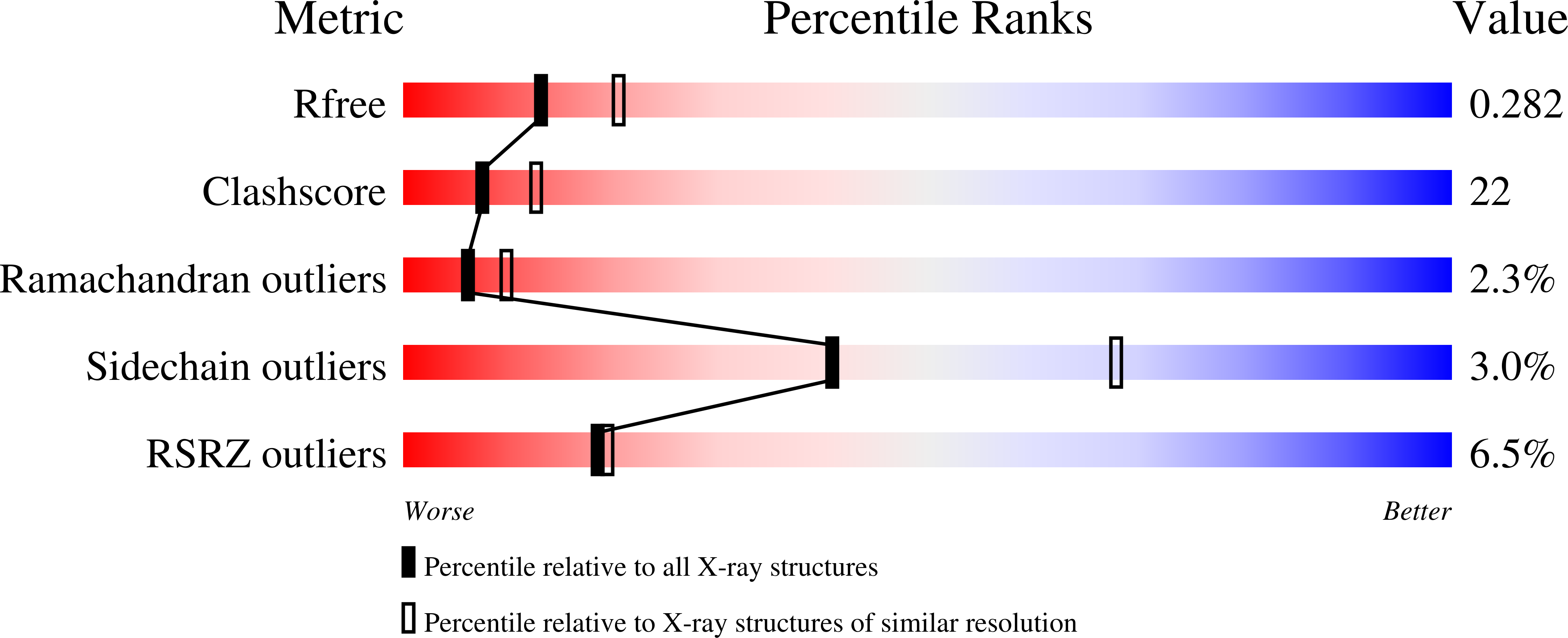
wwPDB Validation   3D Report Full Report


This is version 1.2 of the entry. See complete history


Literature

Crystal structure of E47-NeuroD1/beta2 bHLH domain-DNA complex: heterodimer selectivity and DNA recognition.

Longo, A.Guanga, G.P.Rose, R.B.

(2008) Biochemistry 47: 218-229

  • DOI: https://doi.org/10.1021/bi701527r
  • Primary Citation of Related Structures:  
    2QL2

  • PubMed Abstract: 

    The ubiquitous class I basic helix-loop-helix (bHLH) factor E47 forms heterodimers with multiple tissue specific class II bHLH proteins to regulate distinct differentiation pathways. In order to define how class I- class II heterodimer partners are selected, we determined the crystal structure of the E47-NeuroD1-bHLH dimer in complex with the insulin promoter E-box sequence. Purification of the bHLH domain of E47-NeuroD1 indicates that E47 heterodimers are stable in solution. The interactions between E47 and NeuroD1 in the heterodimer are comparable to the interactions between E47 monomers in the homodimer, including hydrogen bonding, buried hydrophobic surface, and packing interactions. This is consistent with a model in which E47-NeuroD1 heterodimers are favored due to the instability of NeuroD1 homodimers. Although E47-NeuroD1 is oriented uniquely on the E-box sequence (CATCTG) within the promoter of the insulin gene, no direct contacts are observed with the central base pairs within this E-box sequence. We propose that concerted domain motions allow E47 to form specific base contacts in solution. NeuroD1 is restrained from adopting the same base contacts by an additional phosphate backbone interaction by the neurogenic-specific residue His115. Orienting E47-NeuroD1 on promoters may foster protein-protein contacts essential to initiate transcription.


  • Organizational Affiliation

    Department of Molecular and Structural Biochemistry, North Carolina State University, 128 Polk Hall, Raleigh, North Carolina 27695-7622, USA.


Macromolecules

Find similar proteins by:  (by identity cutoff)  |  3D Structure
Entity ID: 1
MoleculeChains Sequence LengthOrganismDetailsImage
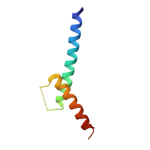
Transcription factor E2-alpha
A, C
60Mus musculusMutation(s): 0 
Gene Names: Tcf3
UniProt
Find proteins for P15806 (Mus musculus)
Explore P15806 
Go to UniProtKB:  P15806
Entity Groups  
Sequence Clusters30% Identity50% Identity70% Identity90% Identity95% Identity100% Identity
UniProt GroupP15806
Sequence Annotations
Expand
  • Reference Sequence
Find similar proteins by:  (by identity cutoff)  |  3D Structure
Entity ID: 2
MoleculeChains Sequence LengthOrganismDetailsImage
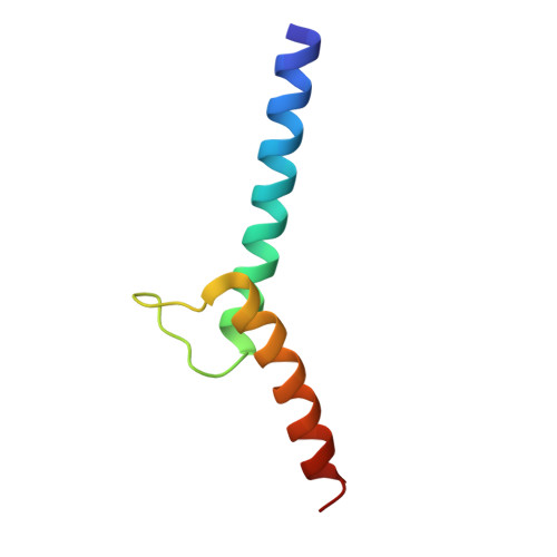
Neurogenic differentiation factor 1
B, D
60Mus musculusMutation(s): 0 
Gene Names: Neurod1Neurod
UniProt
Find proteins for Q60867 (Mus musculus)
Explore Q60867 
Go to UniProtKB:  Q60867
Entity Groups  
Sequence Clusters30% Identity50% Identity70% Identity90% Identity95% Identity100% Identity
UniProt GroupQ60867
Sequence Annotations
Expand
  • Reference Sequence

Find similar nucleic acids by:  Sequence   |   3D Structure  

Entity ID: 3
MoleculeChains LengthOrganismImage
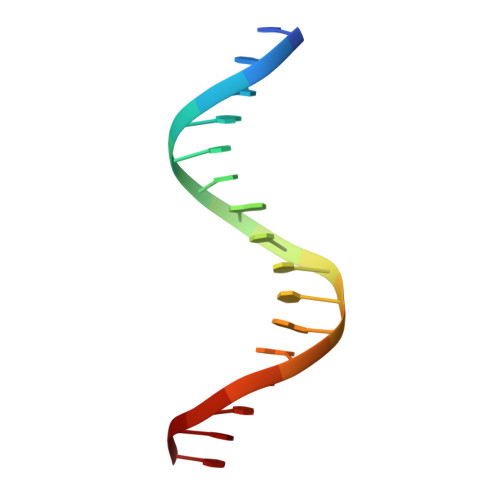
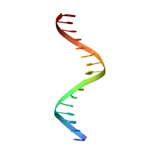
DNA (5'-D(*DTP*DAP*DGP*DGP*DCP*DCP*DAP*DTP*DCP*DTP*DGP*DGP*DTP*DCP*DCP*DT)-3')
E, G
16N/A
Sequence Annotations
Expand
  • Reference Sequence

Find similar nucleic acids by:  Sequence   |   3D Structure  

Entity ID: 4
MoleculeChains LengthOrganismImage
DNA (5'-D(*DAP*DGP*DGP*DAP*DCP*DCP*DAP*DGP*DAP*DTP*DGP*DGP*DCP*DCP*DTP*DA)-3')
F, H
16N/A
Sequence Annotations
Expand
  • Reference Sequence
Experimental Data & Validation

Experimental Data

  • Method: X-RAY DIFFRACTION
  • Resolution: 2.50 Å
  • R-Value Free: 0.289 
  • R-Value Work: 0.248 
  • Space Group: P 21 21 2
Unit Cell:
Length ( Å )Angle ( ˚ )
a = 60.31α = 90
b = 169.54β = 90
c = 52.24γ = 90
Software Package:
Software NamePurpose
SCALAdata processing
CNSrefinement
PDB_EXTRACTdata extraction
AUTOMARdata collection
MOSFLMdata reduction
SCALAdata scaling
CNSphasing

Structure Validation

View Full Validation Report


Entry History 

Deposition Data

Revision History  (Full details and data files)

  • Version 1.0: 2008-11-04
    Type: Initial release
  • Version 1.1: 2011-07-13
    Changes: Version format compliance
  • Version 1.2: 2023-08-30
    Changes: Data collection, Database references, Refinement description