The Partly Folded Back Solution Structure Arrangement of the 30 SCR Domains in Human Complement Receptor Type 1 (CR1) Permits Access to its C3b and C4b Ligands
Furtado, P.B., Huang, C.Y., Ihyembe, D., Hammond, R.A., Marsh, H.C., Perkins, S.J.(2008) J Mol Biol 375: 102-118
- PubMed: 18028942
- DOI: https://doi.org/10.1016/j.jmb.2007.09.085
- Primary Citation of Related Structures:
2Q7Z - PubMed Abstract:
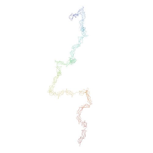
Human complement receptor type 1 (CR1, CD35) is a type I membrane-bound glycoprotein that belongs to the regulators of complement activity (RCA) family. The extra-cellular component of CR1 is comprised of 30 short complement regulator (SCR) domains, whereas complement receptor type 2 (CR2) has 15 SCR domains and factor H (FH) has 20 SCR domains. The domain arrangement of a soluble form of CR1 (sCR1) was studied by X-ray scattering and analytical ultracentrifugation. The radius of gyration R(G) of sCR1 of 13.4(+/-1.1) nm is not much greater than those for CR2 and FH, and its R(G)/R(0) anisotropy ratio is 3.76, compared to ratios of 3.67 for FH and 4.1 for CR2. Unlike CR2, but similar to FH, two cross-sectional R(G) ranges were identified that gave R(XS) values of 4.7(+/-0.2) nm and 1.2(+/-0.7) nm, respectively, showing that the SCR domains adopt a range of conformations including folded-back ones. The distance distribution function P(r) showed that the most commonly occurring distance in sCR1 is at 11.5 nm. Its maximum length of 55 nm is less than double those for CR2 or FH, even though sCR1 has twice the number of SCR domains compared to CR2 Sedimentation equilibrium experiments gave a mean molecular weight of 235 kDa for sCR1. This is consistent with the value of 245 kDa calculated from its composition including 14 N-linked oligosaccharide sites, and confirmed that sCR1 is a monomer in solution. Sedimentation velocity experiments gave a sedimentation coefficient of 5.8 S. From this, the frictional ratio (f/f(0)) of sCR1 was calculated to be 2.29, which is greater than those of 1.96 for CR2 and 1.77 for FH. The constrained scattering modelling of the sCR1 solution structure starting from homologous SCR domain structures generated 5000 trial conformationally randomised models, 43 of which gave good scattering fits to show that sCR1 has a partly folded-back structure. We conclude that the inter-SCR linkers show structural features in common with those in FH, but differ from those in CR2, and the SCR arrangement in CR1 will permit C3b or C4b to access all three ligand sites.
Organizational Affiliation:
Department of Biochemistry and Molecular Biology, University College London, Gower Street, London WC1E 6BT, UK.