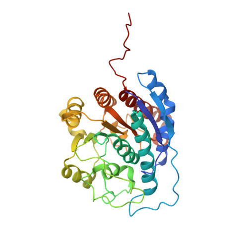
2PHO

Crystal structure of human arginase I complexed with thiosemicarbazide at 1.95 resolution


Experimental Data Snapshot

  • Method: X-RAY DIFFRACTION
  • Resolution: 1.95 Å
  • R-Value Free: 0.219 
  • R-Value Work: 0.169 

wwPDB Validation   3D Report Full Report


This is version 1.3 of the entry. See complete history


Literature

Crystal structure of human arginase I complexed with thiosemicarbazide reveals an unusual thiocarbonyl mu-sulfide ligand in the binuclear manganese cluster.

Di Costanzo, L.Pique, M.E.Christianson, D.W.

(2007) J Am Chem Soc 129: 6388-6389

  • DOI: https://doi.org/10.1021/ja071567j
  • Primary Citation of Related Structures:  
    2PHA, 2PHO, 2ZAV

  • PubMed Abstract: 

    The crystal structure of the human arginase I-thiosemicarbazide complex reveals an unusual thiocarbonyl μ-sulfide ligand in the binuclear manganese cluster. The C=S moiety of thiosemicarbazide bridges Mn 2+ A and Mn 2+ B with coordination distances of 2.6 Å and 2.4 Å, respectively. Otherwise, the binding of thiosemicarbazide to human arginase I does not cause any significant structural changes in the active site. The crystal structure of the unliganded enzyme reveals a hydrogen bonded water molecule that could support proton transfer between a μ-water molecule and H141 to regenerate the nucleophilic μ-hydroxide ion in the final step of catalysis.


  • Organizational Affiliation

    Roy and Diana Vagelos Laboratories, Department of Chemistry, University of Pennsylvania, Philadelphia, PA 19104-6323, USA.


Macromolecules
Find similar proteins by:  (by identity cutoff)  |  3D Structure
Entity ID: 1
MoleculeChains Sequence LengthOrganismDetailsImage
Arginase-1
A, B
322Homo sapiensMutation(s): 0 
Gene Names: ARG1
EC: 3.5.3.1
UniProt & NIH Common Fund Data Resources
Find proteins for P05089 (Homo sapiens)
Explore P05089 
Go to UniProtKB:  P05089
PHAROS:  P05089
GTEx:  ENSG00000118520 
Entity Groups  
Sequence Clusters30% Identity50% Identity70% Identity90% Identity95% Identity100% Identity
UniProt GroupP05089
Sequence Annotations
Expand
  • Reference Sequence
Experimental Data & Validation

Experimental Data

  • Method: X-RAY DIFFRACTION
  • Resolution: 1.95 Å
  • R-Value Free: 0.219 
  • R-Value Work: 0.169 
  • Space Group: P 3
Unit Cell:
Length ( Å )Angle ( ˚ )
a = 90.61α = 90
b = 90.61β = 90
c = 69.63γ = 120
Software Package:
Software NamePurpose
ADSCdata collection
PHASERphasing
CNSrefinement
HKL-2000data reduction
SCALEPACKdata scaling

Structure Validation

View Full Validation Report


Entry History 

Deposition Data

Revision History  (Full details and data files)

  • Version 1.0: 2007-05-08
    Type: Initial release
  • Version 1.1: 2007-12-17
    Changes: Version format compliance
  • Version 1.2: 2011-07-13
    Changes: Derived calculations, Version format compliance
  • Version 1.3: 2023-08-30
    Changes: Data collection, Database references, Derived calculations, Refinement description