2P2R

Crystal structure of the third KH domain of human Poly(C)-Binding Protein-2 in complex with C-rich strand of human telomeric DNA


Experimental Data Snapshot

  • Method: X-RAY DIFFRACTION
  • Resolution: 1.60 Å
  • R-Value Free: 0.242 
  • R-Value Work: 0.205 
  • R-Value Observed: 0.207 

wwPDB Validation   3D Report Full Report


This is version 1.4 of the entry. See complete history


Literature

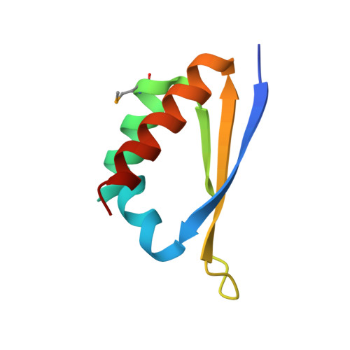
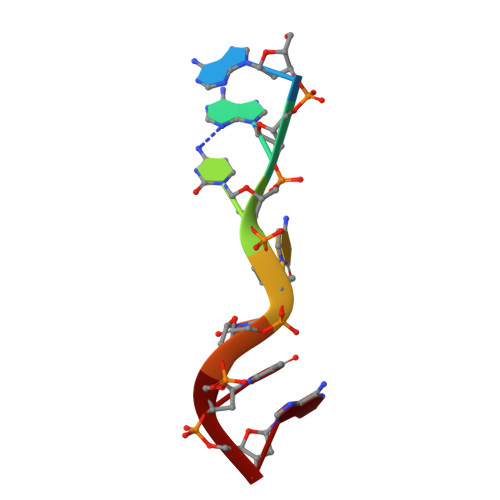
Crystal structure of the third KH domain of human poly(C)-binding protein-2 in complex with a C-rich strand of human telomeric DNA at 1.6 A resolution.

Fenn, S.Du, Z.Lee, J.K.Tjhen, R.Stroud, R.M.James, T.L.

(2007) Nucleic Acids Res 35: 2651-2660

  • DOI: https://doi.org/10.1093/nar/gkm139
  • Primary Citation of Related Structures:  
    2P2R

  • PubMed Abstract: 

    KH (hnRNP K homology) domains, consisting of approximately 70 amino acid residues, are present in a variety of nucleic-acid-binding proteins. Among these are poly(C)-binding proteins (PCBPs), which are important regulators of mRNA stability and posttranscriptional regulation in general. All PCBPs contain three different KH domains and recognize poly(C)-sequences with high affinity and specificity. To reveal the molecular basis of poly(C)-sequence recognition, we have determined the crystal structure, at 1.6 A resolution, of PCBP2 KH3 domain in complex with a 7-nt DNA sequence (5'-AACCCTA-3') corresponding to one repeat of the C-rich strand of human telomeric DNA. The domain assumes a type-I KH fold in a betaalphaalphabetabetaalpha configuration. The protein-DNA interface could be studied in unprecedented detail and is made up of a series of direct and water-mediated hydrogen bonds between the protein and the DNA, revealing an especially dense network involving several structural water molecules for the last 2 nt in the core recognition sequence. Unlike published KH domain structures, the protein crystallizes without protein-protein contacts, yielding new insights into the dimerization properties of different KH domains. A nucleotide platform, an interesting feature found in some RNA molecules, was identified, evidently for the first time in DNA.


  • Organizational Affiliation

    Department of Pharmaceutical Chemistry, University of California, San Francisco, California 94143-2280, USA.


Macromolecules

Find similar proteins by:  (by identity cutoff)  |  3D Structure
Entity ID: 2
MoleculeChains Sequence LengthOrganismDetailsImage
Poly(rC)-binding protein 2B [auth A]76Homo sapiensMutation(s): 1 
Gene Names: PCBP2
UniProt & NIH Common Fund Data Resources
Find proteins for Q15366 (Homo sapiens)
Explore Q15366 
Go to UniProtKB:  Q15366
PHAROS:  Q15366
GTEx:  ENSG00000197111 
Entity Groups  
Sequence Clusters30% Identity50% Identity70% Identity90% Identity95% Identity100% Identity
UniProt GroupQ15366
Sequence Annotations
Expand
  • Reference Sequence

Find similar nucleic acids by:  Sequence   |   3D Structure  

Entity ID: 1
MoleculeChains LengthOrganismImage
C-rich strand of human telomeric DNAA [auth B]7N/A
Sequence Annotations
Expand
  • Reference Sequence
Small Molecules
Ligands 1 Unique
IDChains Name / Formula / InChI Key2D Diagram3D Interactions
CYT
Query on CYT

Download Ideal Coordinates CCD File 
C [auth A]6-AMINOPYRIMIDIN-2(1H)-ONE
C4 H5 N3 O
OPTASPLRGRRNAP-UHFFFAOYSA-N
Modified Residues  1 Unique
IDChains TypeFormula2D DiagramParent
MSE
Query on MSE
B [auth A]L-PEPTIDE LINKINGC5 H11 N O2 SeMET
Experimental Data & Validation

Experimental Data

  • Method: X-RAY DIFFRACTION
  • Resolution: 1.60 Å
  • R-Value Free: 0.242 
  • R-Value Work: 0.205 
  • R-Value Observed: 0.207 
  • Space Group: H 3 2
Unit Cell:
Length ( Å )Angle ( ˚ )
a = 81.069α = 90
b = 81.069β = 90
c = 87.815γ = 120
Software Package:
Software NamePurpose
REFMACrefinement
SCALEPACKdata scaling
CNSphasing
CNSrefinement
DENZOdata reduction
PDB_EXTRACTdata extraction
HKL-2000data reduction
HKL-2000data scaling

Structure Validation

View Full Validation Report


Entry History 

Deposition Data

Revision History  (Full details and data files)

  • Version 1.0: 2007-06-12
    Type: Initial release
  • Version 1.1: 2007-10-16
    Changes: Version format compliance
  • Version 1.2: 2011-07-13
    Changes: Version format compliance
  • Version 1.3: 2017-10-18
    Changes: Advisory, Refinement description
  • Version 1.4: 2019-07-24
    Changes: Data collection, Derived calculations, Refinement description