Structural features of the ligand binding site on human complement protein C8gamma: A member of the lipocalin family
Chiswell, B., Lovelace, L.L., Brannen, C., Ortlund, E.A., Lebioda, L., Sodetz, J.M.(2007) Biochim Biophys Acta 1774: 637-644
- PubMed: 17452033
- DOI: https://doi.org/10.1016/j.bbapap.2007.03.004
- Primary Citation of Related Structures:
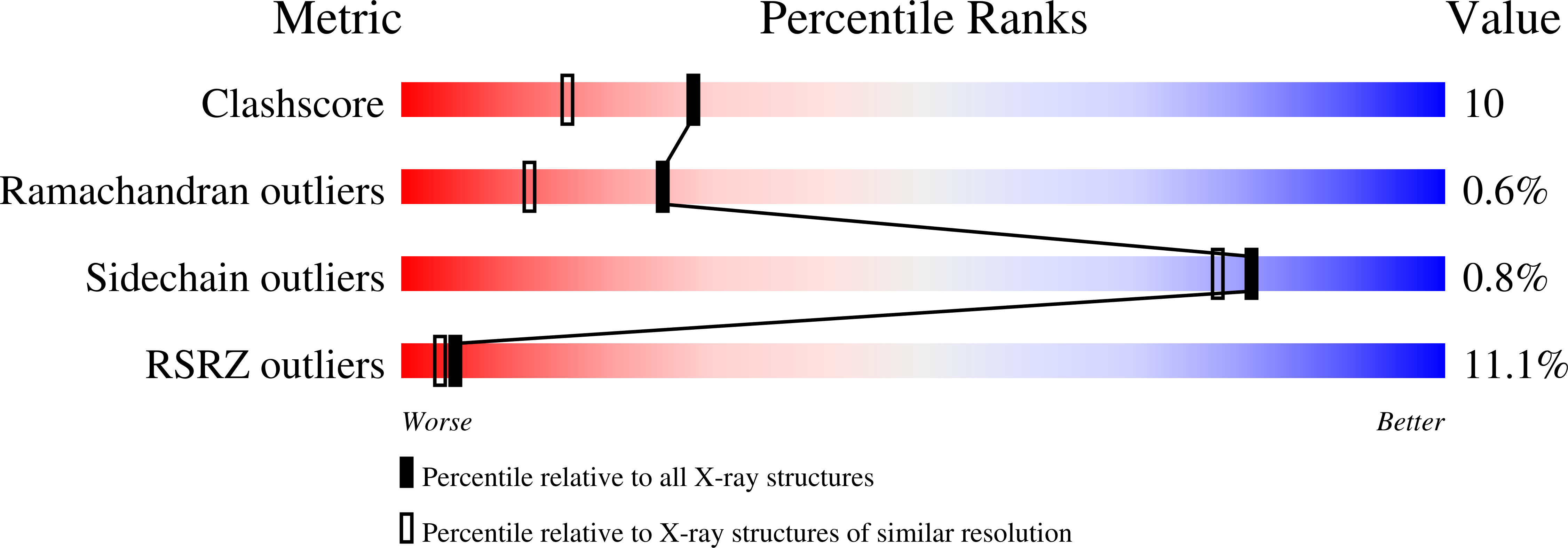
2OVA, 2OVD, 2OVE - PubMed Abstract:
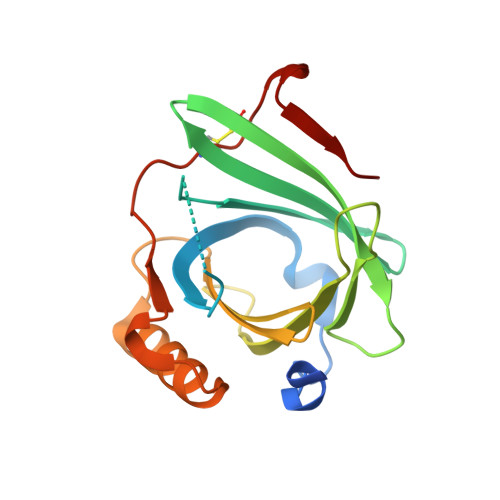
Human C8 is one of five components of the cytolytic membrane attack complex of complement. It contains three subunits (C8alpha, C8beta, C8gamma) arranged as a disulfide-linked C8alpha-gamma heterodimer that is noncovalently associated with C8beta. C8gamma has the distinction of being the only lipocalin in the complement system. Lipocalins have a core beta-barrel structure forming a calyx with a binding site for a small hydrophobic ligand. A natural ligand for C8gamma has not been identified; however previous structural studies indicate C8gamma has a typical lipocalin fold that is suggestive of a ligand-binding capability. A distinctive feature of C8gamma is the division of its putative ligand binding pocket into a hydrophilic upper portion and a large hydrophobic lower cavity. Access to the latter is restricted by the close proximity of two tyrosine side chains (Y83 and Y131). In the present study, binding experiments were performed using lauric acid as a pseudoligand to investigate the potential accessibility of the lower cavity. The crystal structure of a C8gamma.laurate complex revealed that Y83 and Y131 can move to allow penetration of the hydrocarbon chain of laurate into the lower cavity. Introducing a Y83W mutation blocked access but had no effect on the ability of C8gamma to enhance C8 cytolytic activity. Together, these results indicate that the lower cavity in C8gamma could accommodate a ligand if such a ligand has a narrow hydrophobic moiety at one end. Entry of that moiety into the lower cavity would require movement of Y83 and Y131, which act as a gate at the cavity entrance.
Organizational Affiliation:
Department of Chemistry and Biochemistry and School of Medicine, University of South Carolina, Columbia, SC 29208, USA.