2O6V

Crystal structure and solution NMR studies of Lys48-linked tetraubiquitin at neutral pH


Experimental Data Snapshot

  • Method: X-RAY DIFFRACTION
  • Resolution: 2.20 Å
  • R-Value Free: 0.262 
  • R-Value Work: 0.222 
  • R-Value Observed: 0.240 

wwPDB Validation   3D Report Full Report


This is version 1.5 of the entry. See complete history


Literature

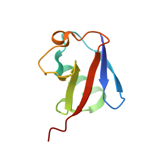
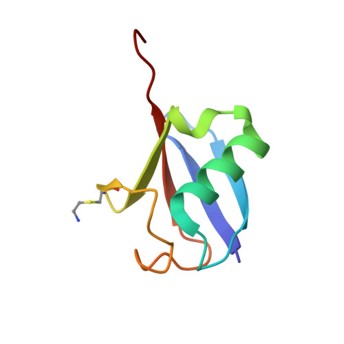
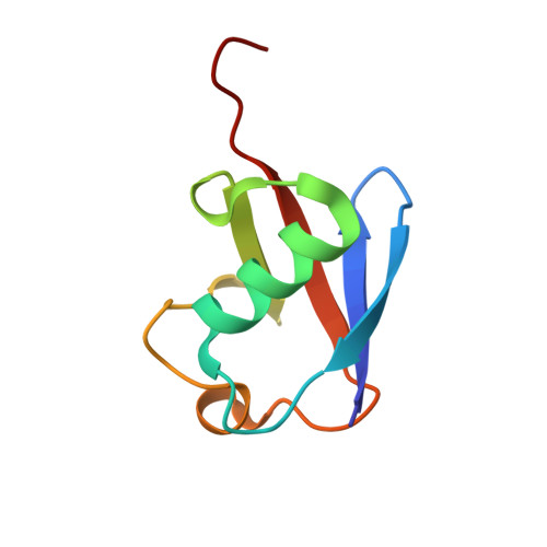
Crystal Structure and Solution NMR Studies of Lys48-linked Tetraubiquitin at Neutral pH

Eddins, M.J.Varadan, R.Fushman, D.Pickart, C.M.Wolberger, C.

(2007) J Mol Biol 367: 204-211

  • DOI: https://doi.org/10.1016/j.jmb.2006.12.065
  • Primary Citation of Related Structures:  
    2O6V

  • PubMed Abstract: 

    Ubiquitin modification of proteins is used as a signal in many cellular processes. Lysine side-chains can be modified by a single ubiquitin or by a polyubiquitin chain, which is defined by an isopeptide bond between the C terminus of one ubiquitin and a specific lysine in a neighboring ubiquitin. Polyubiquitin conformations that result from different lysine linkages presumably differentiate their roles and ability to bind specific targets and enzymes. However, conflicting results have been obtained regarding the precise conformation of Lys48-linked tetraubiquitin. We report the crystal structure of Lys48-linked tetraubiquitin at near-neutral pH. The two tetraubiquitin complexes in the asymmetric unit show the complete connectivity of the chain and the molecular details of the interactions. This tetraubiquitin conformation is consistent with our NMR data as well as with previous studies of diubiquitin and tetraubiquitin in solution at neutral pH. The structure provides a basis for understanding Lys48-linked polyubiquitin recognition under physiological conditions.


  • Organizational Affiliation

    Department of Biophysics and Biophysical Chemistry and the Howard Hughes Medical Institute, Johns Hopkins University School of Medicine, Baltimore, MD 21205, USA.


Macromolecules
Find similar proteins by:  (by identity cutoff)  |  3D Structure
Entity ID: 1
MoleculeChains Sequence LengthOrganismDetailsImage
Ubiquitin
A, C, E, G
76Homo sapiensMutation(s): 0 
UniProt & NIH Common Fund Data Resources
Find proteins for P0CG48 (Homo sapiens)
Explore P0CG48 
Go to UniProtKB:  P0CG48
PHAROS:  P0CG48
GTEx:  ENSG00000150991 
Entity Groups  
Sequence Clusters30% Identity50% Identity70% Identity90% Identity95% Identity100% Identity
UniProt GroupP0CG48
Sequence Annotations
Expand
  • Reference Sequence
Find similar proteins by:  (by identity cutoff)  |  3D Structure
Entity ID: 2
MoleculeChains Sequence LengthOrganismDetailsImage
Ubiquitin
B, F
76Homo sapiensMutation(s): 2 
UniProt & NIH Common Fund Data Resources
Find proteins for P0CG48 (Homo sapiens)
Explore P0CG48 
Go to UniProtKB:  P0CG48
PHAROS:  P0CG48
GTEx:  ENSG00000150991 
Entity Groups  
Sequence Clusters30% Identity50% Identity70% Identity90% Identity95% Identity100% Identity
UniProt GroupP0CG48
Sequence Annotations
Expand
  • Reference Sequence
Find similar proteins by:  (by identity cutoff)  |  3D Structure
Entity ID: 3
MoleculeChains Sequence LengthOrganismDetailsImage
Ubiquitin
D, H
76Homo sapiensMutation(s): 2 
UniProt & NIH Common Fund Data Resources
Find proteins for P0CG48 (Homo sapiens)
Explore P0CG48 
Go to UniProtKB:  P0CG48
PHAROS:  P0CG48
GTEx:  ENSG00000150991 
Entity Groups  
Sequence Clusters30% Identity50% Identity70% Identity90% Identity95% Identity100% Identity
UniProt GroupP0CG48
Sequence Annotations
Expand
  • Reference Sequence
Experimental Data & Validation

Experimental Data

  • Method: X-RAY DIFFRACTION
  • Resolution: 2.20 Å
  • R-Value Free: 0.262 
  • R-Value Work: 0.222 
  • R-Value Observed: 0.240 
  • Space Group: C 1 2 1
Unit Cell:
Length ( Å )Angle ( ˚ )
a = 59.1α = 90
b = 77.08β = 90.32
c = 139.36γ = 90
Software Package:
Software NamePurpose
ADSCdata collection
MOLREPphasing
CNSrefinement
HKL-2000data scaling

Structure Validation

View Full Validation Report


Entry History 

Deposition Data

Revision History  (Full details and data files)

  • Version 1.0: 2007-02-13
    Type: Initial release
  • Version 1.1: 2008-05-01
    Changes: Version format compliance
  • Version 1.2: 2011-07-13
    Changes: Derived calculations, Version format compliance
  • Version 1.3: 2012-06-27
    Changes: Structure summary
  • Version 1.4: 2021-10-20
    Changes: Database references, Derived calculations
  • Version 1.5: 2023-08-30
    Changes: Data collection, Refinement description