2O02

Phosphorylation independent interactions between 14-3-3 and Exoenzyme S: from structure to pathogenesis


Experimental Data Snapshot

  • Method: X-RAY DIFFRACTION
  • Resolution: 1.50 Å
  • R-Value Free: 0.212 
  • R-Value Work: 0.147 
  • R-Value Observed: 0.151 

wwPDB Validation   3D Report Full Report


This is version 1.3 of the entry. See complete history


Literature

Phosphorylation-independent interaction between 14-3-3 and exoenzyme S: from structure to pathogenesis

Ottmann, C.Yasmin, L.Weyand, M.Veesenmeyer, J.L.Diaz, M.H.Palmer, R.H.Francis, M.S.Hauser, A.R.Wittinghofer, A.Hallberg, B.

(2007) EMBO J 26: 902-913

  • DOI: https://doi.org/10.1038/sj.emboj.7601530
  • Primary Citation of Related Structures:  
    2O02

  • PubMed Abstract: 

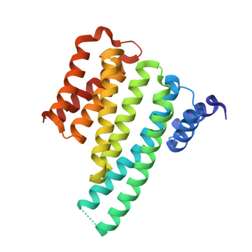
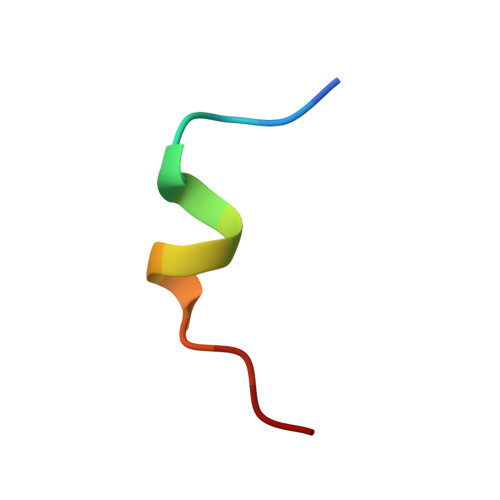
    14-3-3 proteins are phosphoserine/phosphothreonine-recognizing adapter proteins that regulate the activity of a vast array of targets. There are also examples of 14-3-3 proteins binding their targets via unphosphorylated motifs. Here we present a structural and biological investigation of the phosphorylation-independent interaction between 14-3-3 and exoenzyme S (ExoS), an ADP-ribosyltransferase toxin of Pseudomonas aeruginosa. ExoS binds to 14-3-3 in a novel binding mode mostly relying on hydrophobic contacts. The 1.5 A crystal structure is supported by cytotoxicity analysis, which reveals that substitution of the corresponding hydrophobic residues significantly weakens the ability of ExoS to modify the endogenous targets RAS/RAP1 and to induce cell death. Furthermore, mutation of key residues within the ExoS binding site for 14-3-3 impairs virulence in a mouse pneumonia model. In conclusion, we show that ExoS binds 14-3-3 in a novel reversed orientation that is primarily dependent on hydrophobic residues. This interaction is phosphorylation independent and is required for the function of ExoS.


  • Organizational Affiliation

    Department of Structural Biology, Max-Planck-Institute for Molecular Physiology, Dortmund, Germany.


Macromolecules
Find similar proteins by:  (by identity cutoff)  |  3D Structure
Entity ID: 1
MoleculeChains Sequence LengthOrganismDetailsImage
14-3-3 protein zeta/delta
A, B
230Homo sapiensMutation(s): 0 
Gene Names: YWHAZ
UniProt & NIH Common Fund Data Resources
Find proteins for P63104 (Homo sapiens)
Explore P63104 
Go to UniProtKB:  P63104
PHAROS:  P63104
GTEx:  ENSG00000164924 
Entity Groups  
Sequence Clusters30% Identity50% Identity70% Identity90% Identity95% Identity100% Identity
UniProt GroupP63104
Sequence Annotations
Expand
  • Reference Sequence

Find similar proteins by:  Sequence   |   3D Structure  

Entity ID: 2
MoleculeChains Sequence LengthOrganismDetailsImage
ExoS (416-430) peptideC [auth P],
D [auth Q]
14N/AMutation(s): 0 
Entity Groups  
Sequence Clusters30% Identity50% Identity70% Identity90% Identity95% Identity100% Identity
Sequence Annotations
Expand
  • Reference Sequence
Experimental Data & Validation

Experimental Data

  • Method: X-RAY DIFFRACTION
  • Resolution: 1.50 Å
  • R-Value Free: 0.212 
  • R-Value Work: 0.147 
  • R-Value Observed: 0.151 
  • Space Group: P 21 21 21
Unit Cell:
Length ( Å )Angle ( ˚ )
a = 71.33α = 90
b = 72.238β = 90
c = 125.664γ = 90
Software Package:
Software NamePurpose
XSCALEdata scaling
MOLREPphasing
REFMACrefinement
PDB_EXTRACTdata extraction
MAR345data collection
XDSdata reduction

Structure Validation

View Full Validation Report


Entry History 

Deposition Data

Revision History  (Full details and data files)

  • Version 1.0: 2007-11-27
    Type: Initial release
  • Version 1.1: 2011-07-13
    Changes: Version format compliance
  • Version 1.2: 2017-10-18
    Changes: Refinement description
  • Version 1.3: 2023-12-27
    Changes: Data collection, Database references, Derived calculations