Characterization of Ehp, a Secreted Complement Inhibitory Protein from Staphylococcus aureus.
Hammel, M., Sfyroera, G., Pyrpassopoulos, S., Ricklin, D., Ramyar, K.X., Pop, M., Jin, Z., Lambris, J.D., Geisbrecht, B.V.(2007) J Biol Chem 282: 30051-30061
- PubMed: 17699522
- DOI: https://doi.org/10.1074/jbc.M704247200
- Primary Citation of Related Structures:
2NOJ - PubMed Abstract:
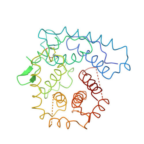
We report here the discovery and characterization of Ehp, a new secreted Staphylococcus aureus protein that potently inhibits the alternative complement activation pathway. Ehp was identified through a genomic scan as an uncharacterized secreted protein from S. aureus, and immunoblotting of conditioned S. aureus culture medium revealed that the Ehp protein was secreted at the highest levels during log-phase bacterial growth. The mature Ehp polypeptide is composed of 80 residues and is 44% identical to the complement inhibitory domain of S. aureus Efb (extracellular fibrinogen-binding protein). We observed preferential binding by Ehp to native and hydrolyzed C3 relative to fully active C3b and found that Ehp formed a subnanomolar affinity complex with these various forms of C3 by binding to its thioester-containing C3d domain. Site-directed mutagenesis demonstrated that Arg(75) and Asn(82) are important in forming the Ehp.C3d complex, but loss of these side chains did not completely disrupt Ehp/C3d binding. This suggested the presence of a second C3d-binding site in Ehp, which was mapped to the proximity of Ehp Asn(63). Further molecular level details of the Ehp/C3d interaction were revealed by solving the 2.7-A crystal structure of an Ehp.C3d complex in which the low affinity site had been mutationally inactivated. Ehp potently inhibited C3b deposition onto sensitized surfaces by the alternative complement activation pathway. This inhibition was directly related to Ehp/C3d binding and was more potent than that seen for Efb-C. An altered conformation in Ehp-bound C3 was detected by monoclonal antibody C3-9, which is specific for a neoantigen exposed in activated forms of C3. Our results suggest that increased inhibitory potency of Ehp relative to Efb-C is derived from the second C3-binding site in this new protein.
Organizational Affiliation:
School of Biological Sciences, University of Missouri-Kansas City, Kansas City, Missouri 64110, USA.