2INT

CRYSTAL STRUCTURE OF RECOMBINANT HUMAN INTERLEUKIN-4


Experimental Data Snapshot

  • Method: X-RAY DIFFRACTION
  • Resolution: 2.35 Å
  • R-Value Work: 0.232 
  • R-Value Observed: 0.232 

wwPDB Validation   3D Report Full Report


This is version 1.4 of the entry. See complete history


Literature

Crystal structure of recombinant human interleukin-4.

Walter, M.R.Cook, W.J.Zhao, B.G.Cameron Jr., R.P.Ealick, S.E.Walter Jr., R.L.Reichert, P.Nagabhushan, T.L.Trotta, P.P.Bugg, C.E.

(1992) J Biol Chem 267: 20371-20376

  • DOI: https://doi.org/10.2210/pdb2int/pdb
  • Primary Citation of Related Structures:  
    2INT

  • PubMed Abstract: 

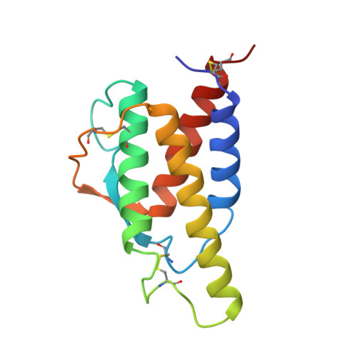
    The crystal structure of recombinant human interleukin-4 (rhuIL-4) was initially determined at 3.5-A resolution by multiple isomorphous replacement techniques and subsequently refined to a resolution of 2.35 A by simulated annealing. The final crystallographic R-factor, based on all data in the range 6.0-2.35 A (7470 reflections), is 0.232. Bond lengths and bond angles in the molecule have root mean square deviations from ideal values of 0.016 A and 2.4 degrees, respectively. The overall structure is highly compact and globular with a predominantly hydrophobic core. The main structural feature of rhuIL-4 is a four alpha-helix bundle, which composes approximately 58% of the structure. The helices are arranged in a left-handed antiparallel bundle with two overhand connections. Within these connections is a two-stranded antiparallel beta-sheet. Both the tertiary and secondary structures of rhuIL-4 are similar to those of human granulocyte-macrophage colony-stimulating factor. Critical regions for receptor binding are proposed.


  • Organizational Affiliation

    Department of Pathology, University of Alabama at Birmingham 35294.


Macromolecules
Find similar proteins by:  (by identity cutoff)  |  3D Structure
Entity ID: 1
MoleculeChains Sequence LengthOrganismDetailsImage
INTERLEUKIN-4129Homo sapiensMutation(s): 0 
UniProt & NIH Common Fund Data Resources
Find proteins for P05112 (Homo sapiens)
Explore P05112 
Go to UniProtKB:  P05112
PHAROS:  P05112
GTEx:  ENSG00000113520 
Entity Groups  
Sequence Clusters30% Identity50% Identity70% Identity90% Identity95% Identity100% Identity
UniProt GroupP05112
Sequence Annotations
Expand
  • Reference Sequence
Experimental Data & Validation

Experimental Data

  • Method: X-RAY DIFFRACTION
  • Resolution: 2.35 Å
  • R-Value Work: 0.232 
  • R-Value Observed: 0.232 
  • Space Group: P 41 21 2
Unit Cell:
Length ( Å )Angle ( ˚ )
a = 91.8α = 90
b = 91.8β = 90
c = 46.2γ = 90
Software Package:
Software NamePurpose
X-PLORmodel building
PROLSQrefinement
X-PLORrefinement
X-PLORphasing

Structure Validation

View Full Validation Report


Entry History 

Revision History  (Full details and data files)

  • Version 1.0: 1994-01-31
    Type: Initial release
  • Version 1.1: 2008-03-24
    Changes: Version format compliance
  • Version 1.2: 2011-07-13
    Changes: Version format compliance
  • Version 1.3: 2019-07-17
    Changes: Data collection, Other, Refinement description
  • Version 1.4: 2019-08-14
    Changes: Data collection, Refinement description