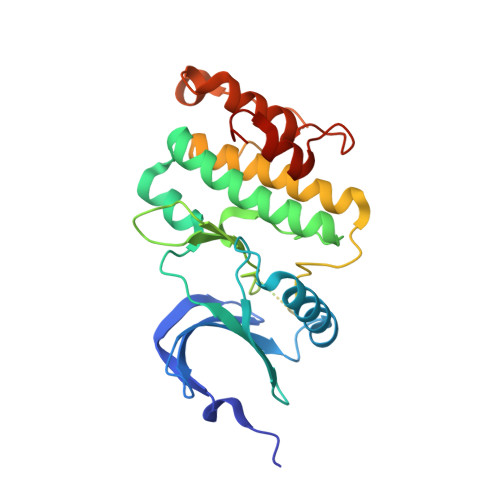
Mitogen-activated protein kinases interacting kinases are autoinhibited by a reprogrammed activation segment.
Jauch, R., Cho, M.K., Netter, C., Schreiter, K., Aicher, B., Zweckstetter, M., Wahl, M.C.(2006) EMBO J 25: 4020-4032
- PubMed: 16917500
- DOI: https://doi.org/10.1038/sj.emboj.7601285
- Primary Citation of Related Structures:
2HW6, 2HW7 - PubMed Abstract:
Autoinhibition is a recurring mode of protein kinase regulation and can be based on diverse molecular mechanisms. Here, we show by crystal structure analysis, nuclear magnetic resonance (NMR)-based nucleotide affinity studies and rational mutagenesis that nonphosphorylated mitogen-activated protein (MAP) kinases interacting kinase (Mnk) 1 is autoinhibited by conversion of the activation segment into an autoinhibitory module. In a Mnk1 crystal structure, the activation segment is repositioned via a Mnk-specific sequence insertion at the N-terminal lobe with the following consequences: (i) the peptide substrate binding site is deconstructed, (ii) the interlobal cleft is narrowed, (iii) an essential Lys-Glu pair is disrupted and (iv) the magnesium-binding loop is locked into an ATP-competitive conformation. Consistently, deletion of the Mnk-specific insertion or removal of a conserved phenylalanine side chain, which induces a blockade of the ATP pocket, increase the ATP affinity of Mnk1. Structural rearrangements required for the activation of Mnks are apparent from the cocrystal structure of a Mnk2 D228G -staurosporine complex and can be modeled on the basis of crystal packing interactions. Our data suggest a novel regulatory mechanism specific for the Mnk subfamily.
Organizational Affiliation:
Max-Planck-Institut für Biophysikalische Chemie, Abteilung Molekulare Entwicklungsbiologie, Göttingen, Germany. jauchr@gis.a-star.edu.sg