2H6G

W102T Protein Farnesyltransferase Mutant Complexed with a Geranylgeranylated DDPTASACVLS Peptide Product at 1.85A Resolution


Experimental Data Snapshot

  • Method: X-RAY DIFFRACTION
  • Resolution: 1.85 Å
  • R-Value Free: 0.207 
  • R-Value Work: 0.187 
  • R-Value Observed: 0.187 

wwPDB Validation   3D Report Full Report


Ligand Structure Quality Assessment 


This is version 2.2 of the entry. See complete history


Literature

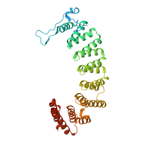
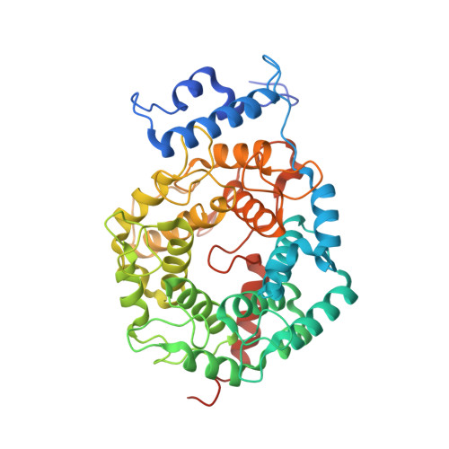
Conversion of protein farnesyltransferase to a geranylgeranyltransferase.

Terry, K.L.Casey, P.J.Beese, L.S.

(2006) Biochemistry 45: 9746-9755

  • DOI: https://doi.org/10.1021/bi060295e
  • Primary Citation of Related Structures:  
    2H6F, 2H6G, 2H6H, 2H6I

  • PubMed Abstract: 

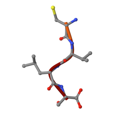
    Posttranslational modifications are essential for the proper function of a number of proteins in the cell. One such modification, the covalent attachment of a single isoprenoid lipid (prenylation), is carried out by the CaaX prenyltransferases, protein farnesyltransferase (FTase) and protein geranylgeranyltransferase type-I (GGTase-I). Substrate proteins of these two enzymes are involved in a variety of cellular functions but are largely associated with signal transduction. These modified proteins include members of the Ras superfamily, heterotrimeric G-proteins, centromeric proteins, and a number of proteins involved in nuclear integrity. Although FTase and GGTase-I are highly homologous, they are quite selective for their substrates, particularly for their isoprenoid diphosphate substrates, FPP and GGPP, respectively. Here, we present both crystallographic and kinetic analyses of mutants designed to explore this isoprenoid specificity and demonstrate that this specificity is dependent upon two enzyme residues in the beta subunits of the enzymes, W102beta and Y365beta in FTase (T49beta and F324beta, respectively, in GGTase-I).


  • Organizational Affiliation

    Department of Biochemistry, Duke University Medical Center, Durham, North Carolina 27710, USA.


Macromolecules
Find similar proteins by:  (by identity cutoff)  |  3D Structure
Entity ID: 1
MoleculeChains Sequence LengthOrganismDetailsImage
Protein farnesyltransferase/geranylgeranyltransferase type I alpha subunit382Homo sapiensMutation(s): 0 
Gene Names: Human FTase alpha subunit
EC: 2.5.1.58 (PDB Primary Data), 2.5.1.59 (PDB Primary Data)
UniProt & NIH Common Fund Data Resources
Find proteins for P49354 (Homo sapiens)
Explore P49354 
Go to UniProtKB:  P49354
PHAROS:  P49354
GTEx:  ENSG00000168522 
Entity Groups  
Sequence Clusters30% Identity50% Identity70% Identity90% Identity95% Identity100% Identity
UniProt GroupP49354
Sequence Annotations
Expand
  • Reference Sequence
Find similar proteins by:  (by identity cutoff)  |  3D Structure
Entity ID: 2
MoleculeChains Sequence LengthOrganismDetailsImage
Protein farnesyltransferase beta subunit437Homo sapiensMutation(s): 1 
Gene Names: Human FTase beta subunit
EC: 2.5.1.58
UniProt & NIH Common Fund Data Resources
Find proteins for P49356 (Homo sapiens)
Explore P49356 
Go to UniProtKB:  P49356
PHAROS:  P49356
GTEx:  ENSG00000257365 
Entity Groups  
Sequence Clusters30% Identity50% Identity70% Identity90% Identity95% Identity100% Identity
UniProt GroupP49356
Sequence Annotations
Expand
  • Reference Sequence

Find similar proteins by:  Sequence   |   3D Structure  

Entity ID: 3
MoleculeChains Sequence LengthOrganismDetailsImage
farnesylated peptideC [auth P]11N/AMutation(s): 0 
Entity Groups  
Sequence Clusters30% Identity50% Identity70% Identity90% Identity95% Identity100% Identity
Sequence Annotations
Expand
  • Reference Sequence
Oligosaccharides

Help

Entity ID: 4
MoleculeChains Length2D Diagram Glycosylation3D Interactions
beta-D-fructofuranose-(2-1)-alpha-D-glucopyranoseD [auth C]2N/A
Glycosylation Resources
GlyTouCan:  G05551OP
GlyCosmos:  G05551OP
Small Molecules
Biologically Interesting Molecules (External Reference) 1 Unique
Experimental Data & Validation

Experimental Data

  • Method: X-RAY DIFFRACTION
  • Resolution: 1.85 Å
  • R-Value Free: 0.207 
  • R-Value Work: 0.187 
  • R-Value Observed: 0.187 
  • Space Group: P 61
Unit Cell:
Length ( Å )Angle ( ˚ )
a = 178.535α = 90
b = 178.535β = 90
c = 64.601γ = 120
Software Package:
Software NamePurpose
HKL-2000data collection
HKL-2000data reduction
CNSrefinement
HKL-2000data scaling
CNSphasing

Structure Validation

View Full Validation Report


Ligand Structure Quality Assessment 


Entry History 

Deposition Data

Revision History  (Full details and data files)

  • Version 1.0: 2006-08-29
    Type: Initial release
  • Version 1.1: 2008-05-01
    Changes: Version format compliance
  • Version 1.2: 2011-07-13
    Changes: Version format compliance
  • Version 2.0: 2020-07-29
    Type: Remediation
    Reason: Carbohydrate remediation
    Changes: Atomic model, Data collection, Database references, Derived calculations, Non-polymer description, Structure summary
  • Version 2.1: 2021-10-20
    Changes: Database references, Structure summary
  • Version 2.2: 2023-08-30
    Changes: Data collection, Refinement description