2GUF

In meso crystal structure of the cobalamin transporter, BtuB


Experimental Data Snapshot

  • Method: X-RAY DIFFRACTION
  • Resolution: 1.95 Å
  • R-Value Free: 0.225 
  • R-Value Work: 0.189 
  • R-Value Observed: 0.191 

wwPDB Validation   3D Report Full Report


Ligand Structure Quality Assessment 


This is version 1.3 of the entry. See complete history


Literature

In Meso Structure of the Cobalamin Transporter, BtuB, at 1.95 A Resolution.

Cherezov, V.Yamashita, E.Liu, W.Zhalnina, M.Cramer, W.A.Caffrey, M.

(2006) J Mol Biol 364: 716-734

  • DOI: https://doi.org/10.1016/j.jmb.2006.09.022
  • Primary Citation of Related Structures:  
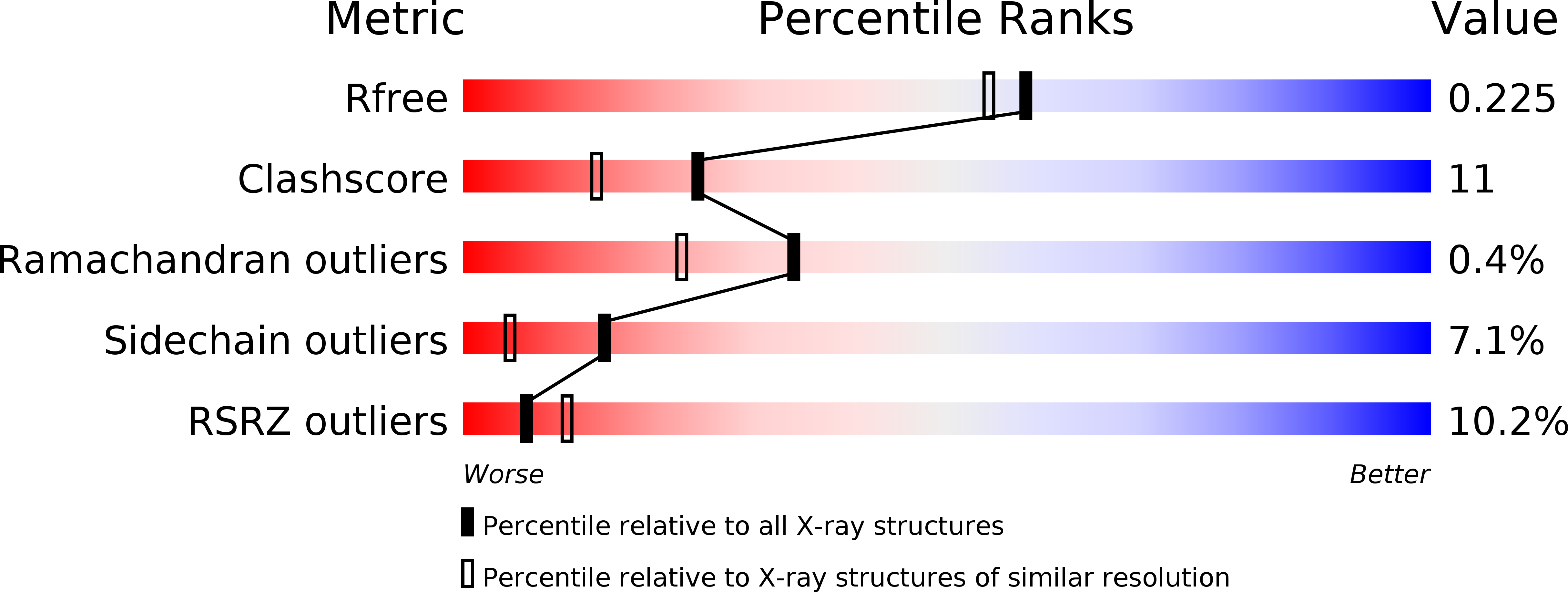
    2GUF

  • PubMed Abstract: 

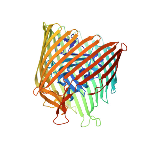
    Crystals of the apo form of the vitamin B12 and colicin receptor, BtuB, that diffract to 1.95 A have been grown by the membrane-based in meso technique. The structure of the protein differs in several details from that of its counterpart grown by the more traditional, detergent-based (in surfo) method. Some of these differences include (i) the five N-terminal residues are resolved in meso, (ii) residues 57-62 in the hatch domain and residues 574-581 in loop 21-22 are disordered in meso and are ordered in surfo, (iii) residues 278-287 in loop 7-8 are resolved in meso, (iv) residues 324-331 in loop 9-10, 396-411 in loop 13-14, 442-458 in loop 15-16 and 526-541 in loop 19-20 have large differences in position between the two crystal forms, as have residues 86-96 in the hatch domain, and (v) the conformation of residues 6 and 7 in the Ton box (considered critical to signal transduction and substrate transport) are entirely different in the two structures. Importantly, the in meso orientation of residues 6 and 7 is similar to that of the vitamin B12-charged state. These data suggest that the "substrate-induced" 180 degrees -rotation of residues 6 and 7 reported in the literature may not be a unique signalling event. The extent to which these findings agree with structural, dynamic and functional insights gleaned from site-directed spin labelling and electron paramagnetic resonance measurements is evaluated. Packing in in meso grown crystals is dense and layered, consistent with the current model for crystallogenesis of membrane proteins in lipidic mesophases. Layered packing has been used to locate the transmembrane hydrophobic surface of the protein. Generally, this is consistent with tryptophan, tyrosine, lipid and CalphaB-factor distributions in the protein, and with predictions based on transfer free energy calculations.


  • Organizational Affiliation

    Department of Chemistry, The Ohio State University, Columbus, OH 43210, USA.


Macromolecules
Find similar proteins by:  (by identity cutoff)  |  3D Structure
Entity ID: 1
MoleculeChains Sequence LengthOrganismDetailsImage
Vitamin B12 transporter btuB594Escherichia coliMutation(s): 0 
Membrane Entity: Yes 
UniProt
Find proteins for P06129 (Escherichia coli (strain K12))
Explore P06129 
Go to UniProtKB:  P06129
Entity Groups  
Sequence Clusters30% Identity50% Identity70% Identity90% Identity95% Identity100% Identity
UniProt GroupP06129
Sequence Annotations
Expand
  • Reference Sequence
Small Molecules
Ligands 3 Unique
IDChains Name / Formula / InChI Key2D Diagram3D Interactions
MPG
Query on MPG

Download Ideal Coordinates CCD File 
B [auth A]
C [auth A]
D [auth A]
E [auth A]
F [auth A]
B [auth A],
C [auth A],
D [auth A],
E [auth A],
F [auth A],
G [auth A],
H [auth A],
I [auth A],
J [auth A],
K [auth A],
L [auth A]
[(Z)-octadec-9-enyl] (2R)-2,3-bis(oxidanyl)propanoate
C21 H40 O4
JPJYKWFFJCWMPK-GDCKJWNLSA-N
MPD
Query on MPD

Download Ideal Coordinates CCD File 
N [auth A],
O [auth A],
P [auth A],
Q [auth A]
(4S)-2-METHYL-2,4-PENTANEDIOL
C6 H14 O2
SVTBMSDMJJWYQN-YFKPBYRVSA-N
FMT
Query on FMT

Download Ideal Coordinates CCD File 
M [auth A]FORMIC ACID
C H2 O2
BDAGIHXWWSANSR-UHFFFAOYSA-N
Experimental Data & Validation

Experimental Data

  • Method: X-RAY DIFFRACTION
  • Resolution: 1.95 Å
  • R-Value Free: 0.225 
  • R-Value Work: 0.189 
  • R-Value Observed: 0.191 
  • Space Group: P 21 21 21
Unit Cell:
Length ( Å )Angle ( ˚ )
a = 72.61α = 90
b = 80.877β = 90
c = 118.681γ = 90
Software Package:
Software NamePurpose
REFMACrefinement
HKL-2000data scaling
MOLREPphasing

Structure Validation

View Full Validation Report


Ligand Structure Quality Assessment 


Entry History 

Deposition Data

Revision History  (Full details and data files)

  • Version 1.0: 2006-12-05
    Type: Initial release
  • Version 1.1: 2008-05-01
    Changes: Version format compliance
  • Version 1.2: 2011-07-13
    Changes: Advisory, Version format compliance
  • Version 1.3: 2024-02-14
    Changes: Data collection, Database references, Derived calculations