2GK2

Crystal structure of the N terminal domain of human CEACAM1


Experimental Data Snapshot

  • Method: X-RAY DIFFRACTION
  • Resolution: 2.20 Å
  • R-Value Free: 0.258 
  • R-Value Work: 0.210 
  • R-Value Observed: 0.213 

wwPDB Validation   3D Report Full Report


This is version 1.4 of the entry. See complete history


Literature

Structure of the N-terminal domain of human CEACAM1: binding target of the opacity proteins during invasion of Neisseria meningitidis and N. gonorrhoeae.

Fedarovich, A.Tomberg, J.Nicholas, R.A.Davies, C.

(2006) Acta Crystallogr D Biol Crystallogr 62: 971-979

  • DOI: https://doi.org/10.1107/S0907444906020737
  • Primary Citation of Related Structures:  
    2GK2

  • PubMed Abstract: 

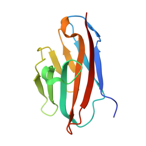
    CEACAM1 is a cellular adhesion molecule whose protein expression is down-regulated in several carcinomas and which also contributes to the pathogenicity of Neisseria by acting as a receptor for Opa proteins. The crystal structure of the N-terminal (D1) domain of human CEACAM1 has been determined at 2.2 Angstrom resolution. The structure shows several differences compared with a lower resolution model of the same domain from mouse solved previously, especially in the functional regions. Mapping of the sites of mutations that lower or abolish the binding of CEACAM1 to Opa proteins shows a distinct clustering of residues on the GFCC'C'' face of the molecule. Prominent amongst these are residues in the C, C' and F strands and the CC' loop. A similar analysis shows that the region responsible for homophilic or heterophilic interactions of CEACAM1 is also on the GFCC'C'' face and overlaps partially with the Opa-binding region. This higher resolution structure of CEACAM1 will facilitate a more precise dissection of its functional regions in the context of neisserial pathogenesis, cellular adhesion and immune evasion.


  • Organizational Affiliation

    Department of Biochemistry and Molecular Biology, Medical University of South Carolina, Charleston, SC 29425, USA.


Macromolecules
Find similar proteins by:  (by identity cutoff)  |  3D Structure
Entity ID: 1
MoleculeChains Sequence LengthOrganismDetailsImage
Carcinoembryonic antigen-related cell adhesion molecule 1
A, B
111Homo sapiensMutation(s): 0 
Gene Names: CEACAM1
UniProt & NIH Common Fund Data Resources
Find proteins for P13688 (Homo sapiens)
Explore P13688 
Go to UniProtKB:  P13688
PHAROS:  P13688
GTEx:  ENSG00000079385 
Entity Groups  
Sequence Clusters30% Identity50% Identity70% Identity90% Identity95% Identity100% Identity
UniProt GroupP13688
Sequence Annotations
Expand
  • Reference Sequence
Experimental Data & Validation

Experimental Data

  • Method: X-RAY DIFFRACTION
  • Resolution: 2.20 Å
  • R-Value Free: 0.258 
  • R-Value Work: 0.210 
  • R-Value Observed: 0.213 
  • Space Group: P 63
Unit Cell:
Length ( Å )Angle ( ˚ )
a = 86.2α = 90
b = 86.2β = 90
c = 62γ = 120
Software Package:
Software NamePurpose
REFMACrefinement
MAR345data collection
HKL-2000data scaling
MOLREPphasing

Structure Validation

View Full Validation Report


Entry History 

Deposition Data

Revision History  (Full details and data files)

  • Version 1.0: 2006-09-05
    Type: Initial release
  • Version 1.1: 2008-05-01
    Changes: Version format compliance
  • Version 1.2: 2011-07-13
    Changes: Advisory, Version format compliance
  • Version 1.3: 2017-10-18
    Changes: Refinement description
  • Version 1.4: 2023-08-30
    Changes: Data collection, Database references, Derived calculations, Refinement description