2EZG

SOLUTION STRUCTURE OF A COMPLEX OF THE THIRD DNA BINDING DOMAIN OF HUMAN HMG-I(Y) BOUND TO DNA DODECAMER CONTAINING THE PRDII SITE OF THE INTERFERON-BETA PROMOTER, NMR, 35 STRUCTURES


Experimental Data Snapshot

  • Method: SOLUTION NMR
  • Conformers Calculated: 35 
  • Conformers Submitted: 35 

wwPDB Validation   3D Report Full Report


This is version 1.3 of the entry. See complete history


Literature

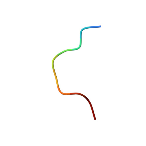
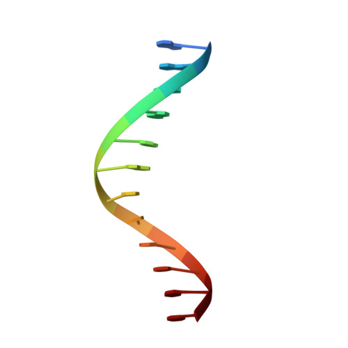
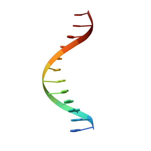
The solution structure of an HMG-I(Y)-DNA complex defines a new architectural minor groove binding motif.

Huth, J.R.Bewley, C.A.Nissen, M.S.Evans, J.N.Reeves, R.Gronenborn, A.M.Clore, G.M.

(1997) Nat Struct Biol 4: 657-665

  • DOI: https://doi.org/10.1038/nsb0897-657
  • Primary Citation of Related Structures:  
    2EZD, 2EZE, 2EZF, 2EZG

  • PubMed Abstract: 

    The solution structure of a complex between a truncated form of HMG-I(Y), consisting of the second and third DNA binding domains (residues 51-90), and a DNA dodecamer containing the PRDII site of the interferon-beta promoter has been solved by multidimensional nuclear magnetic resonance spectroscopy. The stoichiometry of the complex is one molecule of HMG-I(Y) to two molecules of DNA. The structure reveals a new architectural minor groove binding motif which stabilizes B-DNA, thereby facilitating the binding of other transcription factors in the opposing major groove. The interactions involve a central Arg-Gly-Arg motif together with two other modules that participate in extensive hydrophobic and polar contracts. The absence of one of these modules in the third DNA binding domain accounts for its-100 fold reduced affinity relative to the second one.


  • Organizational Affiliation

    Laboratory of Chemical Physics, National Institute of Diabetes and Digestive and Kidney Diseases, National Institutes of Health, Bethesda, Maryland 20892-520, USA.


Macromolecules

Find similar proteins by:  Sequence   |   3D Structure  

Entity ID: 3
MoleculeChains Sequence LengthOrganismDetailsImage
HIGH MOBILITY GROUP PROTEIN HMG-I/HMG-YC [auth A]10Homo sapiensMutation(s): 0 
UniProt & NIH Common Fund Data Resources
Find proteins for P17096 (Homo sapiens)
Explore P17096 
Go to UniProtKB:  P17096
PHAROS:  P17096
GTEx:  ENSG00000137309 
Entity Groups  
Sequence Clusters30% Identity50% Identity70% Identity90% Identity95% Identity100% Identity
UniProt GroupP17096
Sequence Annotations
Expand
  • Reference Sequence

Find similar nucleic acids by:  Sequence   |   3D Structure  

Entity ID: 1
MoleculeChains LengthOrganismImage
DNA (5'-D(*GP*GP*GP*AP*AP*AP*TP*TP*CP*CP*TP*C)-3')A [auth B]12N/A
Sequence Annotations
Expand
  • Reference Sequence

Find similar nucleic acids by:  Sequence   |   3D Structure  

Entity ID: 2
MoleculeChains LengthOrganismImage
DNA (5'-D(*GP*AP*GP*GP*AP*AP*TP*TP*TP*CP*CP*C)-3')B [auth C]12N/A
Sequence Annotations
Expand
  • Reference Sequence
Experimental Data & Validation

Experimental Data

  • Method: SOLUTION NMR
  • Conformers Calculated: 35 
  • Conformers Submitted: 35 

Structure Validation

View Full Validation Report


Entry History 

Deposition Data

Revision History  (Full details and data files)

  • Version 1.0: 1997-10-15
    Type: Initial release
  • Version 1.1: 2008-03-24
    Changes: Version format compliance
  • Version 1.2: 2011-07-13
    Changes: Version format compliance
  • Version 1.3: 2022-03-09
    Changes: Database references, Derived calculations, Other