


Crystal structure of MS0616
Hosaka, T., Nishino, A., Uchikubo, K.-T., Kishishita, S., Murayama, K., Shirouzu, M., Yokoyama, S.To be published.
Experimental Data Snapshot
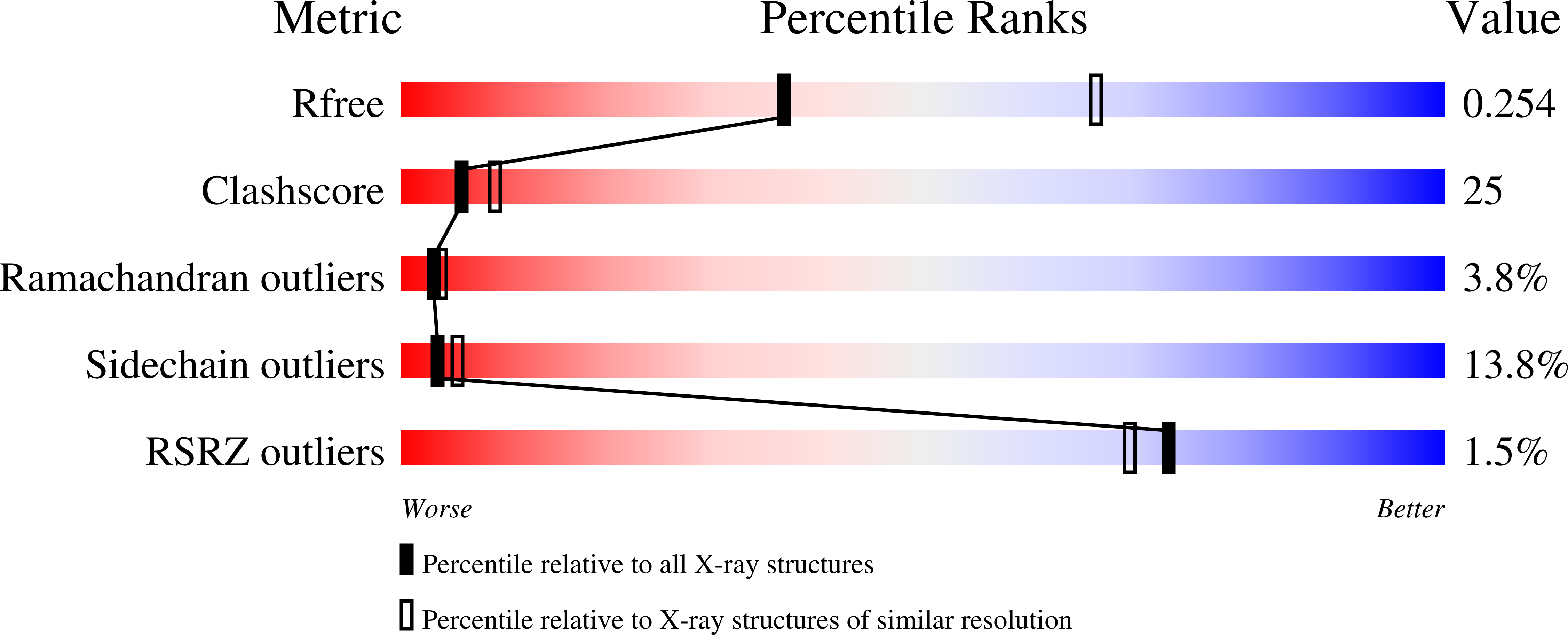
wwPDB Validation 3D Report Full Report
Entity ID: 1 | |||||
|---|---|---|---|---|---|
| Molecule | Chains | Sequence Length | Organism | Details | Image |
| MS0616 | 138 | Mus musculus | Mutation(s): 0 EC: 3.6.1.52 (PDB Primary Data), 3.6.1 (PDB Primary Data) | 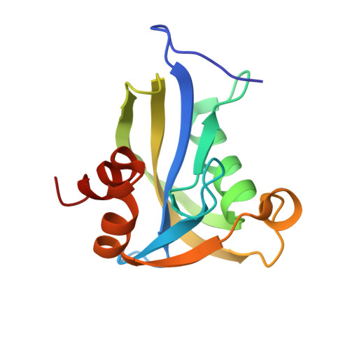 | |
UniProt | |||||
Find proteins for Q8R2U6 (Mus musculus) Explore Q8R2U6 Go to UniProtKB: Q8R2U6 | |||||
Entity Groups | |||||
| Sequence Clusters | 30% Identity50% Identity70% Identity90% Identity95% Identity100% Identity | ||||
| UniProt Group | Q8R2U6 | ||||
Sequence AnnotationsExpand | |||||
| |||||
| Length ( Å ) | Angle ( ˚ ) |
|---|---|
| a = 40.891 | α = 90 |
| b = 41.448 | β = 90 |
| c = 189.288 | γ = 90 |
| Software Name | Purpose |
|---|---|
| REFMAC | refinement |
| HKL-2000 | data reduction |
| HKL-2000 | data scaling |
| CNS | phasing |
RCSB PDB (citation) is hosted by
RCSB PDB is a member of the

