2D7R

Crystal structure of pp-GalNAc-T10 complexed with GalNAc-Ser on lectin domain


Experimental Data Snapshot

  • Method: X-RAY DIFFRACTION
  • Resolution: 2.80 Å
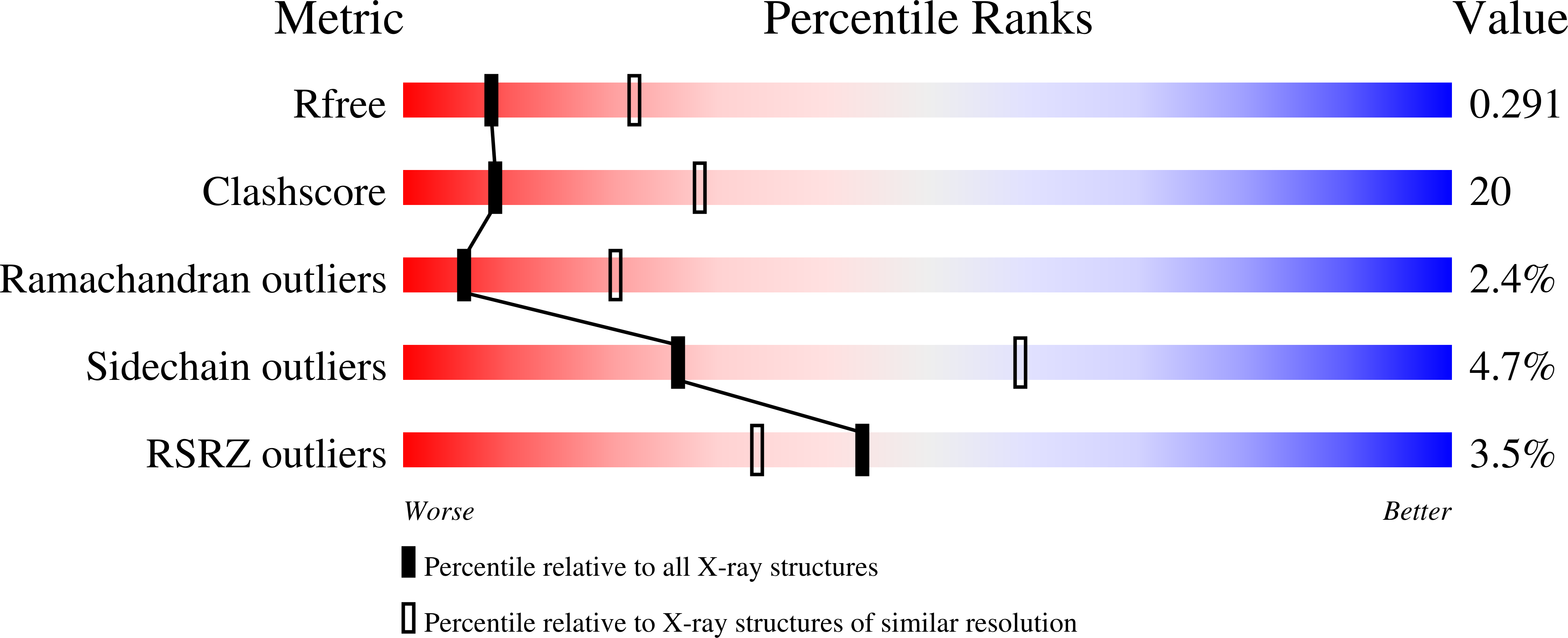
  • R-Value Free: 0.298 
  • R-Value Work: 0.229 
  • R-Value Observed: 0.236 

wwPDB Validation   3D Report Full Report


Ligand Structure Quality Assessment 


This is version 3.1 of the entry. See complete history


Literature

Structural basis of carbohydrate transfer activity by human UDP-GalNAc: polypeptide alpha-N-acetylgalactosaminyltransferase (pp-GalNAc-T10)

Kubota, T.Shiba, T.Sugioka, S.Furukawa, S.Sawaki, H.Kato, R.Wakatsuki, S.Narimatsu, H.

(2006) J Mol Biol 359: 708-727

  • DOI: https://doi.org/10.1016/j.jmb.2006.03.061
  • Primary Citation of Related Structures:  
    2D7I, 2D7R

  • PubMed Abstract: 

    Mucin-type O-glycans are important carbohydrate chains involved in differentiation and malignant transformation. Biosynthesis of the O-glycan is initiated by the transfer of N-acetylgalactosamine (GalNAc) which is catalyzed by UDP-GalNAc:polypeptide alpha-N-acetylgalactosaminyltransferases (pp-GalNAc-Ts). Here we present crystal structures of the pp-GalNAc-T10 isozyme, which has specificity for glycosylated peptides, in complex with the hydrolyzed donor substrate UDP-GalNAc and in complex with GalNAc-serine. A structural comparison with uncomplexed pp-GalNAc-T1 suggests that substantial conformational changes occur in two loops near the catalytic center upon donor substrate binding, and that a distinct interdomain arrangement between the catalytic and lectin domains forms a narrow cleft for acceptor substrates. The distance between the catalytic center and the carbohydrate-binding site on the lectin beta sub-domain influences the position of GalNAc glycosylation on GalNAc-glycosylated peptide substrates. A chimeric enzyme in which the two domains of pp-GalNAc-T10 are connected by a linker from pp-GalNAc-T1 acquires activity toward non-glycosylated acceptors, identifying a potential mechanism for generating the various acceptor specificities in different isozymes to produce a wide range of O-glycans.


  • Organizational Affiliation

    Glycogene Function Team of Research Center for Glycoscience (RCG), National Institute of Advanced Industrial Science and Technology (AIST), Central 2, 1-1-1 Umezono, Tsukuba, Ibaraki 305-8568, Japan.


Macromolecules
Find similar proteins by:  (by identity cutoff)  |  3D Structure
Entity ID: 1
MoleculeChains Sequence LengthOrganismDetailsImage
Polypeptide N-acetylgalactosaminyltransferase 10570Homo sapiensMutation(s): 0 
EC: 2.4.1.41
UniProt & NIH Common Fund Data Resources
Find proteins for Q86SR1 (Homo sapiens)
Explore Q86SR1 
Go to UniProtKB:  Q86SR1
PHAROS:  Q86SR1
GTEx:  ENSG00000164574 
Entity Groups  
Sequence Clusters30% Identity50% Identity70% Identity90% Identity95% Identity100% Identity
UniProt GroupQ86SR1
Sequence Annotations
Expand
  • Reference Sequence
Oligosaccharides

Help

Entity ID: 2
MoleculeChains Length2D Diagram Glycosylation3D Interactions
2-acetamido-2-deoxy-beta-D-glucopyranose-(1-4)-2-acetamido-2-deoxy-beta-D-glucopyranose
B
2N-Glycosylation
Glycosylation Resources
GlyTouCan:  G42666HT
GlyCosmos:  G42666HT
GlyGen:  G42666HT
Small Molecules
Ligands 6 Unique
IDChains Name / Formula / InChI Key2D Diagram3D Interactions
UDP
Query on UDP

Download Ideal Coordinates CCD File 
G [auth A]URIDINE-5'-DIPHOSPHATE
C9 H14 N2 O12 P2
XCCTYIAWTASOJW-XVFCMESISA-N
A2G
Query on A2G

Download Ideal Coordinates CCD File 
I [auth A]2-acetamido-2-deoxy-alpha-D-galactopyranose
C8 H15 N O6
OVRNDRQMDRJTHS-CBQIKETKSA-N
NGA
Query on NGA

Download Ideal Coordinates CCD File 
D [auth A]2-acetamido-2-deoxy-beta-D-galactopyranose
C8 H15 N O6
OVRNDRQMDRJTHS-JAJWTYFOSA-N
NAG
Query on NAG

Download Ideal Coordinates CCD File 
C [auth A],
E [auth A]
2-acetamido-2-deoxy-beta-D-glucopyranose
C8 H15 N O6
OVRNDRQMDRJTHS-FMDGEEDCSA-N
SER
Query on SER

Download Ideal Coordinates CCD File 
H [auth A]SERINE
C3 H7 N O3
MTCFGRXMJLQNBG-REOHCLBHSA-N
MN
Query on MN

Download Ideal Coordinates CCD File 
F [auth A]MANGANESE (II) ION
Mn
WAEMQWOKJMHJLA-UHFFFAOYSA-N
Experimental Data & Validation

Experimental Data

  • Method: X-RAY DIFFRACTION
  • Resolution: 2.80 Å
  • R-Value Free: 0.298 
  • R-Value Work: 0.229 
  • R-Value Observed: 0.236 
  • Space Group: C 2 2 21
Unit Cell:
Length ( Å )Angle ( ˚ )
a = 80.195α = 90
b = 132.812β = 90
c = 138.953γ = 90
Software Package:
Software NamePurpose
HKL-2000data collection
SCALEPACKdata scaling
MOLREPphasing
CNSrefinement
HKL-2000data reduction

Structure Validation

View Full Validation Report


Ligand Structure Quality Assessment 


Entry History 

Deposition Data

Revision History  (Full details and data files)

  • Version 1.0: 2006-11-07
    Type: Initial release
  • Version 1.1: 2008-04-02
    Changes: Version format compliance
  • Version 1.2: 2011-07-13
    Changes: Non-polymer description, Version format compliance
  • Version 2.0: 2020-07-01
    Changes: Atomic model, Data collection, Database references, Derived calculations, Non-polymer description, Structure summary
  • Version 3.0: 2020-07-29
    Type: Remediation
    Reason: Carbohydrate remediation
    Changes: Atomic model, Data collection, Derived calculations, Structure summary
  • Version 3.1: 2023-10-25
    Changes: Data collection, Database references, Refinement description, Structure summary