2A2N

Crystal Structure of the peptidylprolyl isomerase domain of Human PPWD1


Experimental Data Snapshot

  • Method: X-RAY DIFFRACTION
  • Resolution: 1.65 Å
  • R-Value Free: 0.245 
  • R-Value Work: 0.199 
  • R-Value Observed: 0.202 

wwPDB Validation   3D Report Full Report


This is version 1.3 of the entry. See complete history


Literature

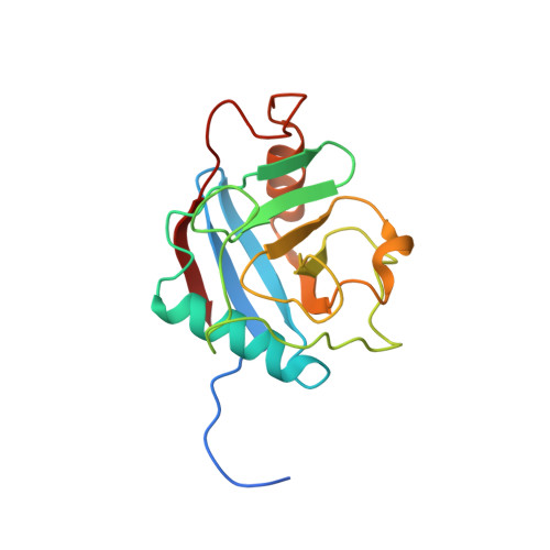
The crystal structure of human WD40 repeat-containing peptidylprolyl isomerase (PPWD1).

Davis, T.L.Walker, J.R.Ouyang, H.MacKenzie, F.Butler-Cole, C.Newman, E.M.Eisenmesser, E.Z.Dhe-Paganon, S.

(2008) FEBS J 275: 2283-2295

  • DOI: https://doi.org/10.1111/j.1742-4658.2008.06381.x
  • Primary Citation of Related Structures:  
    2A2N

  • PubMed Abstract: 

    Cyclophilins comprise one of the three classes of peptidylprolyl isomerases found in all eukaryotic and prokaryotic organisms, as well as viruses. Many of the 17 annotated human cyclophilins contain the catalytic domain in tandem with other domains, and many of the specific functions of a particular cyclophilin or its associated domains remain unknown. The structure of the isomerase domain from a spliceosome-associated cyclophilin, PPWD1 (peptidylprolyl isomerase containing WD40 repeat), has been solved to 1.65 A. In the crystal, the N-terminus of one isomerase domain is bound in the active site of a neighboring isomerase molecule in a manner analogous to substrate. NMR solution studies show that this sequence binds to the active site of the cyclophilin, but cannot be turned over by the enzyme. A pseudo-substrate immediately N-terminal to the cyclophilin domain in PPWD1 could have wider implications for the function of this cyclophilin in the spliceosome, where it is located in human cells.


  • Organizational Affiliation

    Structural Genomics Consortium, Banting Institute, University of Toronto, ON, Canada.


Macromolecules
Find similar proteins by:  (by identity cutoff)  |  3D Structure
Entity ID: 1
MoleculeChains Sequence LengthOrganismDetailsImage
peptidylprolyl isomerase domain and WD repeat containing 1
A, B, C
176Homo sapiensMutation(s): 0 
Gene Names: PPWD1
EC: 5.2.1.8
UniProt & NIH Common Fund Data Resources
Find proteins for Q96BP3 (Homo sapiens)
Explore Q96BP3 
Go to UniProtKB:  Q96BP3
PHAROS:  Q96BP3
GTEx:  ENSG00000113593 
Entity Groups  
Sequence Clusters30% Identity50% Identity70% Identity90% Identity95% Identity100% Identity
UniProt GroupQ96BP3
Sequence Annotations
Expand
  • Reference Sequence
Small Molecules
Ligands 1 Unique
IDChains Name / Formula / InChI Key2D Diagram3D Interactions
GOL
Query on GOL

Download Ideal Coordinates CCD File 
D [auth C]GLYCEROL
C3 H8 O3
PEDCQBHIVMGVHV-UHFFFAOYSA-N
Experimental Data & Validation

Experimental Data

  • Method: X-RAY DIFFRACTION
  • Resolution: 1.65 Å
  • R-Value Free: 0.245 
  • R-Value Work: 0.199 
  • R-Value Observed: 0.202 
  • Space Group: C 1 2 1
Unit Cell:
Length ( Å )Angle ( ˚ )
a = 139.658α = 90
b = 39.893β = 122.33
c = 115.638γ = 90
Software Package:
Software NamePurpose
REFMACrefinement
HKL-2000data reduction
SCALEPACKdata scaling
PHASERphasing

Structure Validation

View Full Validation Report


Entry History 

Revision History  (Full details and data files)

  • Version 1.0: 2005-07-05
    Type: Initial release
  • Version 1.1: 2008-04-30
    Changes: Version format compliance
  • Version 1.2: 2011-07-13
    Changes: Non-polymer description, Version format compliance
  • Version 1.3: 2023-08-23
    Changes: Data collection, Database references, Derived calculations, Refinement description