1ZAK

ADENYLATE KINASE FROM MAIZE IN COMPLEX WITH THE INHIBITOR P1,P5-BIS(ADENOSINE-5'-)PENTAPHOSPHATE (AP5A)


Experimental Data Snapshot

  • Method: X-RAY DIFFRACTION
  • Resolution: 3.50 Å
  • R-Value Work: 0.218 
  • R-Value Observed: 0.218 

wwPDB Validation   3D Report Full Report


Ligand Structure Quality Assessment 


This is version 1.3 of the entry. See complete history


Literature

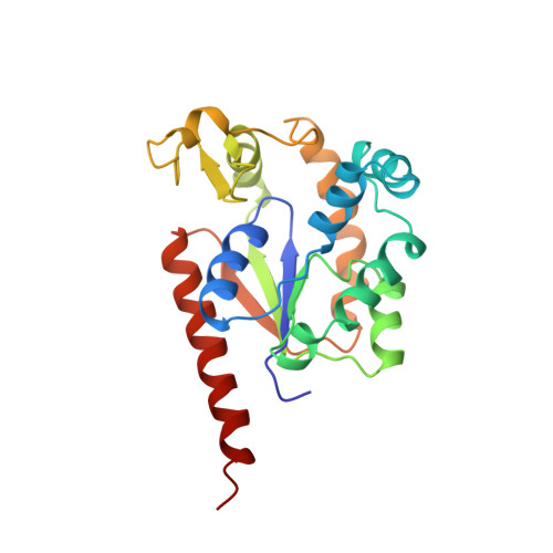
Structure, catalysis and supramolecular assembly of adenylate kinase from maize.

Wild, K.Grafmuller, R.Wagner, E.Schulz, G.E.

(1997) Eur J Biochem 250: 326-331

  • DOI: https://doi.org/10.1111/j.1432-1033.1997.0326a.x
  • Primary Citation of Related Structures:  
    1ZAK

  • PubMed Abstract: 

    The crystal structure of adenylate kinase from maize ligated with an inhibitor has been determined by molecular replacement and refined to 3.5-A resolution. The enzyme keeps the ATP/ADP/AMP equilibrium in the cell. In the C4 plant maize, it has the special task to recycle the AMP produced in large amounts in primary CO2 assimilation. The established structure explains the side reaction with CMP. Moreover, it shows infinite rods that can be readily discerned in the crystal packing. In comparison with homologues, two structural differences that are crucial for this supramolecular assembly are evident. We propose that the rods represent a natural inactive storage form that assembles at night when maize stops CO2 assimilation and thus most of the AMP production in its C4 cycle. The enzyme is particularly abundant in mesophyll chloroplasts, where such an assembly would release appreciable amounts of water that can be used in other processes during the night.


  • Organizational Affiliation

    Institut für Organische Chemie und Biochemie, Albert-Ludwigs-Universität, Freiburg im Breisgau, Germany.


Macromolecules
Find similar proteins by:  (by identity cutoff)  |  3D Structure
Entity ID: 1
MoleculeChains Sequence LengthOrganismDetailsImage
ADENYLATE KINASE
A, B
222Zea maysMutation(s): 0 
EC: 2.7.4.3
UniProt
Find proteins for P43188 (Zea mays)
Explore P43188 
Go to UniProtKB:  P43188
Entity Groups  
Sequence Clusters30% Identity50% Identity70% Identity90% Identity95% Identity100% Identity
UniProt GroupP43188
Sequence Annotations
Expand
  • Reference Sequence
Small Molecules
Ligands 1 Unique
IDChains Name / Formula / InChI Key2D Diagram3D Interactions
AP5
Query on AP5

Download Ideal Coordinates CCD File 
C [auth A],
D [auth B]
BIS(ADENOSINE)-5'-PENTAPHOSPHATE
C20 H29 N10 O22 P5
OIMACDRJUANHTJ-XPWFQUROSA-N
Experimental Data & Validation

Experimental Data

  • Method: X-RAY DIFFRACTION
  • Resolution: 3.50 Å
  • R-Value Work: 0.218 
  • R-Value Observed: 0.218 
  • Space Group: P 3
Unit Cell:
Length ( Å )Angle ( ˚ )
a = 83.3α = 90
b = 83.3β = 90
c = 69.9γ = 120
Software Package:
Software NamePurpose
X-PLORmodel building
X-PLORrefinement
XDSdata reduction
MERGEdata scaling
X-PLORphasing

Structure Validation

View Full Validation Report


Ligand Structure Quality Assessment 


Entry History 

Deposition Data

Revision History  (Full details and data files)

  • Version 1.0: 1998-04-08
    Type: Initial release
  • Version 1.1: 2008-03-24
    Changes: Version format compliance
  • Version 1.2: 2011-07-13
    Changes: Version format compliance
  • Version 1.3: 2023-08-09
    Changes: Database references, Derived calculations, Other, Refinement description