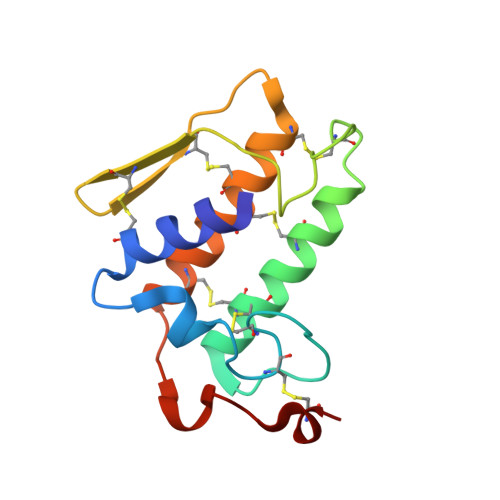
1P7O

Crystal structure of phospholipase A2 (MIPLA4) from Micropechis ikaheka


Experimental Data Snapshot

  • Method: X-RAY DIFFRACTION
  • Resolution: 2.30 Å
  • R-Value Free: 0.274 
  • R-Value Work: 0.220 
  • R-Value Observed: 0.220 

wwPDB Validation   3D Report Full Report


This is version 1.3 of the entry. See complete history


Literature

Structure and function comparison of Micropechis ikaheka snake venom phospholipase A2 isoenzymes

Lok, S.M.Gao, R.Rouault, M.Lambeau, G.Gopalakrishnakone, P.Swaminathan, K.

(2005) FEBS J 272: 1211-1220

  • DOI: https://doi.org/10.1111/j.1742-4658.2005.04547.x
  • Primary Citation of Related Structures:  
    1OZY, 1P7O, 1PWO

  • PubMed Abstract: 

    Comparison of the crystal structures of three Micropechis ikaheka phospholipase A2 isoenzymes (MiPLA2, MiPLA3 and MiPLA4, which exhibit different levels of pharmacological effects) shows that their C-terminus (residues 110-124) is the most variable. M-Type receptor binding affinity of the isoenzymes has also been investigated and MiPLA4 binds to the rabbit M-type receptor with high affinity. Examination of surface charges of the isoenzymes reveals a trend of increase in positive charges with potency. The isoenzymes are shown to oligomerize in a concentration-dependent manner in a semi-denaturing gel. The C-termini of the medium (MiPLA4) and highly potent (MiPLA2) isoenzyme molecules cluster together, forming a highly exposed area. A BLAST search using the sequence of the most potent MiPLA2 results in high similarity to Staphylococcus aureus clotting factor A and cadherin 11. This might explain the myotoxicity, anticoagulant and hemoglobinuria effects of MiPLA2s.


  • Organizational Affiliation

    Institute of Molecular and Cell Biology, Singapore.


Macromolecules
Find similar proteins by:  (by identity cutoff)  |  3D Structure
Entity ID: 1
MoleculeChains Sequence LengthOrganismDetailsImage
PHOSPHOLIPASE A2
A, B, C, D, E
A, B, C, D, E, F
124Micropechis ikahecaMutation(s): 0 
EC: 3.1.1.4
Membrane Entity: Yes 
UniProt
Find proteins for Q45Z29 (Tropidechis carinatus)
Explore Q45Z29 
Go to UniProtKB:  Q45Z29
Entity Groups  
Sequence Clusters30% Identity50% Identity70% Identity90% Identity95% Identity100% Identity
UniProt GroupQ45Z29
Sequence Annotations
Expand
  • Reference Sequence
Experimental Data & Validation

Experimental Data

  • Method: X-RAY DIFFRACTION
  • Resolution: 2.30 Å
  • R-Value Free: 0.274 
  • R-Value Work: 0.220 
  • R-Value Observed: 0.220 
  • Space Group: I 41
Unit Cell:
Length ( Å )Angle ( ˚ )
a = 88.038α = 90
b = 88.038β = 90
c = 262.941γ = 90
Software Package:
Software NamePurpose
CNSrefinement
HKL-2000data reduction
SCALEPACKdata scaling
MOLREPphasing

Structure Validation

View Full Validation Report


Entry History 

Deposition Data

Revision History  (Full details and data files)

  • Version 1.0: 2004-09-28
    Type: Initial release
  • Version 1.1: 2008-04-29
    Changes: Version format compliance
  • Version 1.2: 2011-07-13
    Changes: Derived calculations, Version format compliance
  • Version 1.3: 2024-04-03
    Changes: Data collection, Database references, Refinement description