1P32

CRYSTAL STRUCTURE OF HUMAN P32, A DOUGHNUT-SHAPED ACIDIC MITOCHONDRIAL MATRIX PROTEIN


Experimental Data Snapshot

  • Method: X-RAY DIFFRACTION
  • Resolution: 2.25 Å
  • R-Value Free: 0.234 
  • R-Value Work: 0.173 

wwPDB Validation   3D Report Full Report


This is version 1.5 of the entry. See complete history


Literature

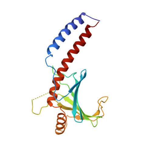
Crystal structure of human p32, a doughnut-shaped acidic mitochondrial matrix protein.

Jiang, J.Zhang, Y.Krainer, A.R.Xu, R.M.

(1999) Proc Natl Acad Sci U S A 96: 3572-3577

  • DOI: https://doi.org/10.1073/pnas.96.7.3572
  • Primary Citation of Related Structures:  
    1P32

  • PubMed Abstract: 

    Human p32 (also known as SF2-associated p32, p32/TAP, and gC1qR) is a conserved eukaryotic protein that localizes predominantly in the mitochondrial matrix. It is thought to be involved in mitochondrial oxidative phosphorylation and in nucleus-mitochondrion interactions. We report the crystal structure of p32 determined at 2.25 A resolution. The structure reveals that p32 adopts a novel fold with seven consecutive antiparallel beta-strands flanked by one N-terminal and two C-terminal alpha-helices. Three monomers form a doughnut-shaped quaternary structure with an unusually asymmetric charge distribution on the surface. The implications of the structure on previously proposed functions of p32 are discussed and new specific functional properties are suggested.


  • Organizational Affiliation

    W. M. Keck Structural Biology Laboratory, Cold Spring Harbor Laboratory, Cold Spring Harbor, NY 11724, USA.


Macromolecules
Find similar proteins by:  (by identity cutoff)  |  3D Structure
Entity ID: 1
MoleculeChains Sequence LengthOrganismDetailsImage
MITOCHONDRIAL MATRIX PROTEIN, SF2P32
A, B, C
209Homo sapiensMutation(s): 1 
UniProt & NIH Common Fund Data Resources
Find proteins for Q07021 (Homo sapiens)
Explore Q07021 
Go to UniProtKB:  Q07021
PHAROS:  Q07021
GTEx:  ENSG00000108561 
Entity Groups  
Sequence Clusters30% Identity50% Identity70% Identity90% Identity95% Identity100% Identity
UniProt GroupQ07021
Sequence Annotations
Expand
  • Reference Sequence
Experimental Data & Validation

Experimental Data

  • Method: X-RAY DIFFRACTION
  • Resolution: 2.25 Å
  • R-Value Free: 0.234 
  • R-Value Work: 0.173 
  • Space Group: P 1 21 1
Unit Cell:
Length ( Å )Angle ( ˚ )
a = 58.625α = 90
b = 56.481β = 95.99
c = 93.831γ = 90
Software Package:
Software NamePurpose
X-PLORmodel building
CNSrefinement
DENZOdata reduction
SCALEPACKdata scaling
X-PLORphasing
CNSphasing

Structure Validation

View Full Validation Report


Entry History 

Deposition Data

Revision History  (Full details and data files)

  • Version 1.0: 1999-04-06
    Type: Initial release
  • Version 1.1: 2008-04-26
    Changes: Version format compliance
  • Version 1.2: 2011-07-13
    Changes: Version format compliance
  • Version 1.3: 2018-03-14
    Changes: Database references
  • Version 1.4: 2018-08-29
    Changes: Data collection, Source and taxonomy, Structure summary
  • Version 1.5: 2023-12-27
    Changes: Data collection, Database references