The domains of protein S from Myxococcus xanthus: structure, stability and interactions.
Wenk, M., Baumgartner, R., Holak, T.A., Huber, R., Jaenicke, R., Mayr, E.M.(1999) J Mol Biol 286: 1533-1545
- PubMed: 10064714
- DOI: https://doi.org/10.1006/jmbi.1999.2582
- Primary Citation of Related Structures:
1NPS - PubMed Abstract:
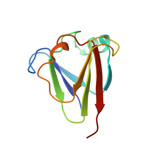
Protein S from Myxococcus xanthus is a member of the beta gamma-crystallin superfamily. Its N and C-terminal domains (NPS and CPS, respectively) show a high degree of structural similarity and possess the capacity to bind two calcium ions per domain. For NPS, their positions were determined by X-ray diffraction at 1.8 A resolution, making use of molecular replacement with the NMR structure as search model. The overall topology of NPS is found to be practically the same as in complete protein S. In natural protein S, the domains fold independently, with a significant increase in stability and cooperativity of folding in the presence of Ca2+. The recombinant isolated domains are stable monomers which do not show any tendency to combine to "nicked" full-length protein S. In order to investigate the stability and folding of natural protein S and its isolated domains, spectroscopic techniques were applied, measuring the reversible urea and temperature-induced unfolding transitions at varying pH. The increment of Ca2+ to the free energy of stabilization amounts to -10 and -5 kJ/mol for NPS and CPS, respectively. For both NPS and CPS, in the absence and in the presence of 3 mM CaCl2, the two-state model is valid. Comparing DeltaGU-->N for CPS (-21 kJ/mol at pH 7, liganded with Ca2+) with its increment in the intact two-domain protein, the stability of the isolated domain turns out to be decreased in a pH-dependent manner. In contrast, the stability of Ca2+-loaded NPS (DeltaGU-->N=-31 kJ/mol, pH 7) is nearly unchanged down to pH 2 where Ca2+ is released (DeltaGU-->N=-26 kJ/mol, pH 2). In intact protein S, the N-terminal domain is destabilized relative to NPS. Evidently, apart from Ca2+ binding, well-defined domain interactions contribute significantly to the overall stability of intact protein S.
Organizational Affiliation:
Institut für Biophysik und Physikalische Biochemie, Universität Regensburg, Regensburg, D-93040, Germany.