1N26

Crystal Structure of the extra-cellular domains of Human Interleukin-6 Receptor alpha chain


Experimental Data Snapshot

  • Method: X-RAY DIFFRACTION
  • Resolution: 2.40 Å
  • R-Value Free: 0.290 
  • R-Value Work: 0.220 
  • R-Value Observed: 0.220 

wwPDB Validation   3D Report Full Report


This is version 2.2 of the entry. See complete history


Literature

Structure of the extracellular domains of the human interleukin-6 receptor alpha-chain.

Varghese, J.N.Moritz, R.L.Lou, M.-Z.van Donkelaar, A.Ji, H.Ivancic, N.Branson, K.M.Hall, N.E.Simpson, R.J.

(2002) Proc Natl Acad Sci U S A 99: 15959-15964

  • DOI: https://doi.org/10.1073/pnas.232432399
  • Primary Citation of Related Structures:  
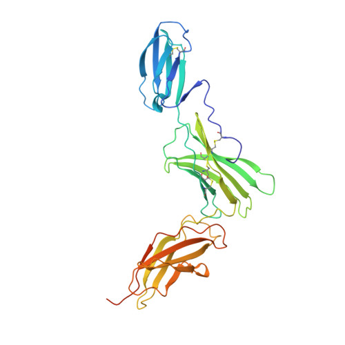
    1N26

  • PubMed Abstract: 

    Dysregulated production of IL-6 and its receptor (IL-6R) are implicated in the pathogenesis of multiple myeloma, autoimmune diseases and prostate cancer. The IL-6R complex comprises two molecules each of IL-6, IL-6R, and the signaling molecule, gp130. Here, we report the x-ray structure (2.4 A) of the IL-6R ectodomains. The N-terminal strand of the Ig-like domain (D(1)) is disulfide-bonded to domain D(2), and domains D(2) and D(3), the cytokine-binding domain, are structurally similar to known cytokine-binding domains. The head-to-tail packing of two closely associated IL-6R molecules observed in the crystal may be representative of the configuration of the physiological dimer of IL-6R and provides new insight into the architecture of the IL-6R complex.


  • Organizational Affiliation

    Biomolecular Research Institute and Commonwealth Scientific and Industrial Research Organization Health Sciences and Nutrition, 343 Royal Parade, Parkville 3052, Australia.


Macromolecules
Find similar proteins by:  (by identity cutoff)  |  3D Structure
Entity ID: 1
MoleculeChains Sequence LengthOrganismDetailsImage
IL-6 Receptor alpha chain325Homo sapiensMutation(s): 0 
Gene Names: IL6R
UniProt & NIH Common Fund Data Resources
Find proteins for P08887 (Homo sapiens)
Explore P08887 
Go to UniProtKB:  P08887
PHAROS:  P08887
GTEx:  ENSG00000160712 
Entity Groups  
Sequence Clusters30% Identity50% Identity70% Identity90% Identity95% Identity100% Identity
UniProt GroupP08887
Sequence Annotations
Expand
  • Reference Sequence
Oligosaccharides

Help

Entity ID: 2
MoleculeChains Length2D Diagram Glycosylation3D Interactions
2-acetamido-2-deoxy-alpha-D-glucopyranose-(1-4)-alpha-D-mannopyranose-(1-6)-beta-D-mannopyranose-(1-4)-2-acetamido-2-deoxy-beta-D-glucopyranose-(1-4)-2-acetamido-2-deoxy-beta-D-glucopyranose
B
5N-Glycosylation
Glycosylation Resources
GlyTouCan:  G31218GY
GlyCosmos:  G31218GY
Entity ID: 3
MoleculeChains Length2D Diagram Glycosylation3D Interactions
2-acetamido-2-deoxy-beta-D-glucopyranose-(1-4)-2-acetamido-2-deoxy-beta-D-glucopyranose
C
2N-Glycosylation
Glycosylation Resources
GlyTouCan:  G42666HT
GlyCosmos:  G42666HT
GlyGen:  G42666HT
Small Molecules
Experimental Data & Validation

Experimental Data

  • Method: X-RAY DIFFRACTION
  • Resolution: 2.40 Å
  • R-Value Free: 0.290 
  • R-Value Work: 0.220 
  • R-Value Observed: 0.220 
  • Space Group: P 43 21 2
Unit Cell:
Length ( Å )Angle ( ˚ )
a = 51.13α = 90
b = 51.13β = 90
c = 303.388γ = 90
Software Package:
Software NamePurpose
DENZOdata reduction
SCALEPACKdata scaling
SHARPphasing
CNSrefinement

Structure Validation

View Full Validation Report


Entry History 

Deposition Data

Revision History  (Full details and data files)

  • Version 1.0: 2002-12-18
    Type: Initial release
  • Version 1.1: 2008-04-28
    Changes: Version format compliance
  • Version 1.2: 2011-07-13
    Changes: Non-polymer description, Version format compliance
  • Version 2.0: 2020-07-29
    Type: Remediation
    Reason: Carbohydrate remediation
    Changes: Advisory, Atomic model, Data collection, Derived calculations, Structure summary
  • Version 2.1: 2023-11-15
    Changes: Advisory, Data collection, Database references, Derived calculations, Structure summary
  • Version 2.2: 2024-04-03
    Changes: Refinement description