Crystal structure of deoxygenated Limulus polyphemus subunit II hemocyanin at 2.18 A resolution: clues for a mechanism for allosteric regulation.
Hazes, B., Magnus, K.A., Bonaventura, C., Bonaventura, J., Dauter, Z., Kalk, K.H., Hol, W.G.(1993) Protein Sci 2: 597-619
- PubMed: 8518732
- DOI: https://doi.org/10.1002/pro.5560020411
- Primary Citation of Related Structures:
1LLA, 1NOL - PubMed Abstract:
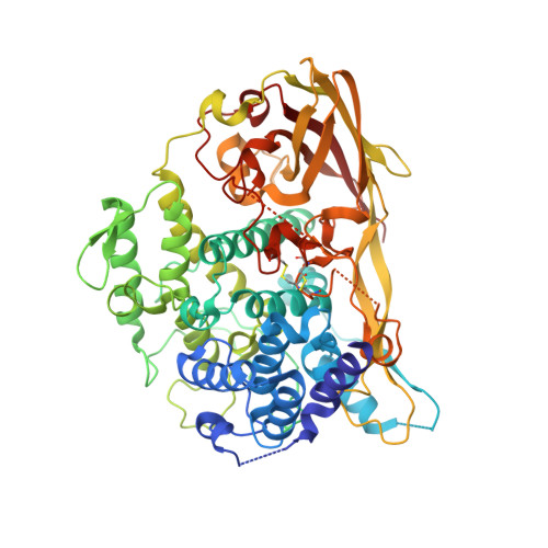
The crystal structure of Limulus polyphemus subunit type II hemocyanin in the deoxygenated state has been determined to a resolution of 2.18 A. Phase information for this first structure of a cheliceratan hemocyanin was obtained by molecular replacement using the crustacean hemocyanin structure of Panulirus interruptus. The most striking observation in the Limulus structure is the unexpectedly large distance of 4.6 A between both copper ions in the oxygen-binding site. Each copper has approximate trigonal planar coordination by three histidine N epsilon atoms. No bridging ligand between the copper ions could be detected. Other important new discoveries are (1) the presence of a cis-peptide bond between Glu 309 and Ser 310, with the carbonyl oxygen of the peptide plane hydrogen bonded to the N delta atom of the copper B ligand His 324; (2) localization of a chloride-binding site in the interface between the first and second domain; (3) localization of a putative calcium-binding site in the third domain. Furthermore, comparison of Limulus versus Panulirus hemocyanin revealed considerable tertiary and quaternary rigid body movements, although the overall folds are similar. Within the subunit, the first domain is rotated by about 7.5 degrees with respect to the other two domains, whereas within the hexamer the major movement is a 3.1 degrees rotation of the trimers with respect to each other. The rigid body rotation of the first domain suggests a structural mechanism for the allosteric regulation by chloride ions and probably causes the cooperative transition of the hexamer between low and high oxygen affinity states. In this postulated mechanism, the fully conserved Phe49 is the key residue that couples conformational changes of the dinuclear copper site into movements of the first domain.
Organizational Affiliation:
BIOSON Research Institute, Department of Chemistry, University of Groningen, The Netherlands.