1HOX

CRYSTAL STRUCTURE OF RABBIT PHOSPHOGLUCOSE ISOMERASE COMPLEXED WITH FRUCTOSE-6-PHOSPHATE


Experimental Data Snapshot

  • Method: X-RAY DIFFRACTION
  • Resolution: 2.10 Å
  • R-Value Free: 0.248 
  • R-Value Work: 0.217 
  • R-Value Observed: 0.221 

wwPDB Validation   3D Report Full Report


Ligand Structure Quality Assessment 


This is version 1.5 of the entry. See complete history


Literature

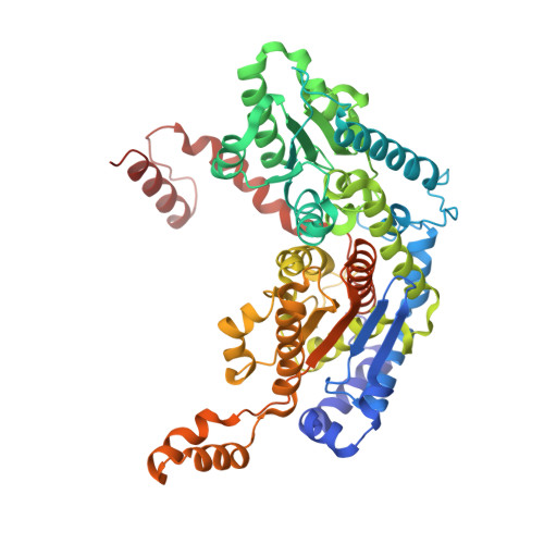
Crystal structure of rabbit phosphoglucose isomerase complexed with its substrate D-fructose 6-phosphate.

Lee, J.H.Chang, K.Z.Patel, V.Jeffery, C.J.

(2001) Biochemistry 40: 7799-7805

  • DOI: https://doi.org/10.1021/bi002916o
  • Primary Citation of Related Structures:  
    1HOX

  • PubMed Abstract: 

    Phosphoglucose isomerase (PGI, EC 5.3.1.9) catalyzes the interconversion of D-glucose 6-phosphate (G6P) and D-fructose 6-phosphate (F6P) and plays important roles in glycolysis and gluconeogenesis. Biochemical characterization of the enzyme has led to a proposed multistep catalytic mechanism. First, the enzyme catalyzes ring opening to yield the open chain form of the substrate. Then isomerization proceeds via proton transfer between C2 and C1 of a cis-enediol(ate) intermediate to yield the open chain form of the product. Catalysis proceeds in both the G6P to F6P and F6P to G6P directions, so both G6P and F6P are substrates. X-ray crystal structure analysis of rabbit and bacterial PGI has previously identified the location of the enzyme active site, and a recent crystal structure of rabbit PGI identified Glu357 as a candidate functional group for transferring the proton. However, it was not clear which active site amino acid residues catalyze the ring opening step. In this paper, we report the X-ray crystal structure of rabbit PGI complexed with the cyclic form of its substrate, D-fructose 6-phosphate, at 2.1 A resolution. The location of the substrate relative to the side chains of His388 suggest that His388 promotes ring opening by protonating the ring oxygen. Glu216 helps to position His388, and a water molecule that is held in position by Lys518 and Thr214 accepts a proton from the hydroxyl group at C2. Comparison to a structure of rabbit PGI with 5PAA bound indicates that ring opening is followed by loss of the protonated water molecule and conformational changes in the substrate and the protein so that a helix containing amino acids 513-520 moves in toward the substrate to form additional hydrogen bonds with the substrate.


  • Organizational Affiliation

    Laboratory for Molecular Biology, MC567, Department of Biology, University of Illinois at Chicago, Chicago, Illinois 60607, USA.


Macromolecules
Find similar proteins by:  (by identity cutoff)  |  3D Structure
Entity ID: 1
MoleculeChains Sequence LengthOrganismDetailsImage
PHOSPHOGLUCOSE ISOMERASE
A, B
558Oryctolagus cuniculusMutation(s): 0 
EC: 5.3.1.9
UniProt
Find proteins for Q9N1E2 (Oryctolagus cuniculus)
Explore Q9N1E2 
Go to UniProtKB:  Q9N1E2
Entity Groups  
Sequence Clusters30% Identity50% Identity70% Identity90% Identity95% Identity100% Identity
UniProt GroupQ9N1E2
Sequence Annotations
Expand
  • Reference Sequence
Small Molecules
Ligands 1 Unique
IDChains Name / Formula / InChI Key2D Diagram3D Interactions
F6P
Query on F6P

Download Ideal Coordinates CCD File 
C [auth A],
D [auth B]
6-O-phosphono-beta-D-fructofuranose
C6 H13 O9 P
BGWGXPAPYGQALX-ARQDHWQXSA-N
Experimental Data & Validation

Experimental Data

  • Method: X-RAY DIFFRACTION
  • Resolution: 2.10 Å
  • R-Value Free: 0.248 
  • R-Value Work: 0.217 
  • R-Value Observed: 0.221 
  • Space Group: C 2 2 21
Unit Cell:
Length ( Å )Angle ( ˚ )
a = 82.438α = 90
b = 119.429β = 90
c = 272.137γ = 90
Software Package:
Software NamePurpose
CNSrefinement
DENZOdata reduction
SCALEPACKdata scaling
CNSphasing

Structure Validation

View Full Validation Report


Ligand Structure Quality Assessment 


Entry History 

Deposition Data

Revision History  (Full details and data files)

  • Version 1.0: 2001-07-20
    Type: Initial release
  • Version 1.1: 2008-04-27
    Changes: Version format compliance
  • Version 1.2: 2011-07-13
    Changes: Version format compliance
  • Version 1.3: 2011-11-16
    Changes: Atomic model
  • Version 1.4: 2020-07-29
    Type: Remediation
    Reason: Carbohydrate remediation
    Changes: Data collection, Derived calculations, Structure summary
  • Version 1.5: 2023-08-09
    Changes: Data collection, Database references, Refinement description, Structure summary