1DO2

TRIGONAL CRYSTAL FORM OF HEAT SHOCK LOCUS U (HSLU) FROM ESCHERICHIA COLI


Experimental Data Snapshot

  • Method: X-RAY DIFFRACTION
  • Resolution: 4.00 Å
  • R-Value Free: 0.285 
  • R-Value Work: 0.229 
  • R-Value Observed: 0.229 

wwPDB Validation   3D Report Full Report


This is version 1.4 of the entry. See complete history


Literature

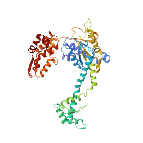
The structures of HsIU and the ATP-dependent protease HsIU-HsIV.

Bochtler, M.Hartmann, C.Song, H.K.Bourenkov, G.P.Bartunik, H.D.Huber, R.

(2000) Nature 403: 800-805

  • DOI: https://doi.org/10.1038/35001629
  • Primary Citation of Related Structures:  
    1DO0, 1DO2

  • PubMed Abstract: 

    The degradation of cytoplasmic proteins is an ATP-dependent process. Substrates are targeted to a single soluble protease, the 26S proteasome, in eukaryotes and to a number of unrelated proteases in prokaryotes. A surprising link emerged with the discovery of the ATP-dependent protease HslVU (heat shock locus VU) in Escherichia coli. Its protease component HslV shares approximately 20% sequence similarity and a conserved fold with 20S proteasome beta-subunits. HslU is a member of the Hsp100 (Clp) family of ATPases. Here we report the crystal structures of free HslU and an 820,000 relative molecular mass complex of HslU and HslV-the first structure of a complete set of components of an ATP-dependent protease. HslV and HslU display sixfold symmetry, ruling out mechanisms of protease activation that require a symmetry mismatch between the two components. Instead, there is conformational flexibility and domain motion in HslU and a localized order-disorder transition in HslV. Individual subunits of HslU contain two globular domains in relative orientations that correlate with nucleotide bound and unbound states. They are surprisingly similar to their counterparts in N-ethylmaleimide-sensitive fusion protein, the prototype of an AAA-ATPase. A third, mostly alpha-helical domain in HslU mediates the contact with HslV and may be the structural equivalent of the amino-terminal domains in proteasomal AAA-ATPases.


  • Organizational Affiliation

    Max-Planck-Institut für Biochemie, Planegg, Germany.


Macromolecules
Find similar proteins by:  (by identity cutoff)  |  3D Structure
Entity ID: 1
MoleculeChains Sequence LengthOrganismDetailsImage
PROTEIN (HEAT SHOCK LOCUS U)
A, B, C, D
442Escherichia coliMutation(s): 0 
UniProt
Find proteins for P0A6H5 (Escherichia coli (strain K12))
Explore P0A6H5 
Go to UniProtKB:  P0A6H5
Entity Groups  
Sequence Clusters30% Identity50% Identity70% Identity90% Identity95% Identity100% Identity
UniProt GroupP0A6H5
Sequence Annotations
Expand
  • Reference Sequence
Small Molecules
Ligands 1 Unique
IDChains Name / Formula / InChI Key2D Diagram3D Interactions
ANP
Query on ANP

Download Ideal Coordinates CCD File 
E [auth A],
F [auth C]
PHOSPHOAMINOPHOSPHONIC ACID-ADENYLATE ESTER
C10 H17 N6 O12 P3
PVKSNHVPLWYQGJ-KQYNXXCUSA-N
Experimental Data & Validation

Experimental Data

  • Method: X-RAY DIFFRACTION
  • Resolution: 4.00 Å
  • R-Value Free: 0.285 
  • R-Value Work: 0.229 
  • R-Value Observed: 0.229 
  • Space Group: P 3 2 1
Unit Cell:
Length ( Å )Angle ( ˚ )
a = 201.777α = 90
b = 201.777β = 90
c = 171.628γ = 120
Software Package:
Software NamePurpose
AMoREphasing
X-PLORrefinement
DENZOdata reduction
SCALEPACKdata scaling

Structure Validation

View Full Validation Report


Entry History 

Deposition Data

Revision History  (Full details and data files)

  • Version 1.0: 2000-02-18
    Type: Initial release
  • Version 1.1: 2008-04-27
    Changes: Version format compliance
  • Version 1.2: 2011-07-13
    Changes: Derived calculations, Version format compliance
  • Version 1.3: 2024-02-07
    Changes: Data collection, Database references, Derived calculations
  • Version 1.4: 2024-04-03
    Changes: Refinement description