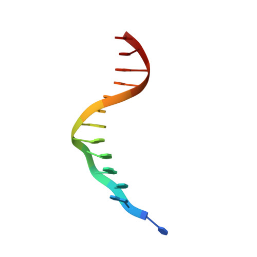
Three-dimensional structures of bulge-containing DNA fragments.
Joshua-Tor, L., Frolow, F., Appella, E., Hope, H., Rabinovich, D., Sussman, J.L.(1992) J Mol Biol 225: 397-431
- PubMed: 1593627
- DOI: https://doi.org/10.1016/0022-2836(92)90929-e
- Primary Citation of Related Structures:
1D31 - PubMed Abstract:
The three-dimensional structure of a DNA tridecamer d(CGCAGAATTCGCG)2 containing bulged adenine bases was determined by single crystal X-ray diffraction methods, at 120 K, to 2.6 A resolution. The structure is a B-DNA type double helix with a single duplex in the asymmetric unit. One of the bulged adenine bases loops out from the double helix, while the other stacks in to it. This is in contrast to our preliminary finding, which indicated that both adenine bases were looped out. This revised model was confirmed by the use of a covalently bound heavy-atom derivative. The conformation of the looped-out bulge hardly disrupts base stacking interactions of the bases flanking it. This is achieved by the backbone making a "loop-the-loop" curve with the extra adenine flipping over with respect to the other nucleotides in the strand. The looped-out base intercalates into the stacked-in bulge site of a symmetrically related duplex. The looped-out and stacked-in bases form an A.A reversed Hoogsteen base-pair that stacks between the surrounding base-pairs, thus stabilizing both bulges. The double helix is frayed at one end with the two "melted" bases participating in intermolecular interactions. A related structure, of the same tridecamer, after soaking the crystals with proflavin, was determined to 3.2 A resolution. The main features of this B-DNA duplex are basically similar to the native tridecamer but differ in detail especially in the conformation of the bulged-out base. Accommodation of a large perturbation such as that described here with minimal disruption of the double helix shows both the flexibility and resiliency of the DNA molecule.
Organizational Affiliation:
Department of Structural Biology, Weizmann Institute of Science, Rehovot, Israel.