1UUH

Hyaluronan binding domain of human CD44


Experimental Data Snapshot

  • Method: X-RAY DIFFRACTION
  • Resolution: 2.20 Å
  • R-Value Free: 0.249 
  • R-Value Work: 0.186 

wwPDB Validation   3D Report Full Report


This is version 1.3 of the entry. See complete history


Literature

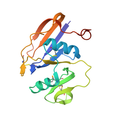
Structure of the Regulatory Hyaluronan-Binding Domain in the Inflammatory Leukocyte Homing Receptor Cd44

Teriete, P.Banerji, S.Noble, M.Blundell, C.Wright, A.Pickford, A.Lowe, E.Mahoney, D.Tammi, M.Kahmann, J.Campbell, I.Day, A.Jackson, D.

(2004) Mol Cell 13: 483

  • DOI: https://doi.org/10.1016/s1097-2765(04)00080-2
  • Primary Citation of Related Structures:  
    1POZ, 1UUH

  • PubMed Abstract: 

    Adhesive interactions involving CD44, the cell surface receptor for hyaluronan, underlie fundamental processes such as inflammatory leukocyte homing and tumor metastasis. Regulation of such events is critical and appears to be effected by changes in CD44 N-glycosylation that switch the receptor "on" or "off" under appropriate circumstances. How altered glycosylation influences binding of hyaluronan to the lectin-like Link module in CD44 is unclear, although evidence suggests additional flanking sequences peculiar to CD44 may be involved. Here we show using X-ray crystallography and NMR spectroscopy that these sequences form a lobular extension to the Link module, creating an enlarged HA binding domain and a formerly unidentified protein fold. Moreover, the disposition of key N-glycosylation sites reveals how specific sugar chains could alter both the affinity and avidity of CD44 HA binding. Our results provide the necessary structural framework for understanding the diverse functions of CD44 and developing novel therapeutic strategies.


  • Organizational Affiliation

    Medical Research Council Human Immunology Unit, Weatherall Institute of Molecular Medicine, John Radcliffe Hospital, Headington, Oxford OX3 9DS, United Kingdom.


Macromolecules
Find similar proteins by:  (by identity cutoff)  |  3D Structure
Entity ID: 1
MoleculeChains Sequence LengthOrganismDetailsImage
CD44 ANTIGEN
A, B
159Homo sapiensMutation(s): 0 
UniProt & NIH Common Fund Data Resources
Find proteins for P16070 (Homo sapiens)
Explore P16070 
Go to UniProtKB:  P16070
PHAROS:  P16070
GTEx:  ENSG00000026508 
Entity Groups  
Sequence Clusters30% Identity50% Identity70% Identity90% Identity95% Identity100% Identity
UniProt GroupP16070
Sequence Annotations
Expand
  • Reference Sequence
Small Molecules
Modified Residues  1 Unique
IDChains TypeFormula2D DiagramParent
MSE
Query on MSE
A, B
L-PEPTIDE LINKINGC5 H11 N O2 SeMET
Experimental Data & Validation

Experimental Data

  • Method: X-RAY DIFFRACTION
  • Resolution: 2.20 Å
  • R-Value Free: 0.249 
  • R-Value Work: 0.186 
  • Space Group: P 21 21 21
Unit Cell:
Length ( Å )Angle ( ˚ )
a = 48.881α = 90
b = 77.266β = 90
c = 87.668γ = 90
Software Package:
Software NamePurpose
REFMACrefinement
SOLVE/RESOLVEphasing

Structure Validation

View Full Validation Report


Entry History 

Deposition Data

Revision History  (Full details and data files)

  • Version 1.0: 2004-03-04
    Type: Initial release
  • Version 1.1: 2011-05-08
    Changes: Version format compliance
  • Version 1.2: 2011-07-13
    Changes: Version format compliance
  • Version 1.3: 2019-05-29
    Changes: Data collection, Derived calculations, Experimental preparation, Other