1UL1

Crystal structure of the human FEN1-PCNA complex


Experimental Data Snapshot

  • Method: X-RAY DIFFRACTION
  • Resolution: 2.90 Å
  • R-Value Free: 0.284 
  • R-Value Work: 0.220 

wwPDB Validation   3D Report Full Report


This is version 1.3 of the entry. See complete history


Literature

Structural basis for recruitment of human flap endonuclease 1 to PCNA

Sakurai, S.Kitano, K.Yamaguchi, H.Okada, K.Hamada, K.Fukuda, K.Uchida, M.Ohtsuka, E.Morioka, H.Hakoshima, T.

(2005) EMBO J 24: 683-693

  • DOI: https://doi.org/10.1038/sj.emboj.7600519
  • Primary Citation of Related Structures:  
    1UL1

  • PubMed Abstract: 

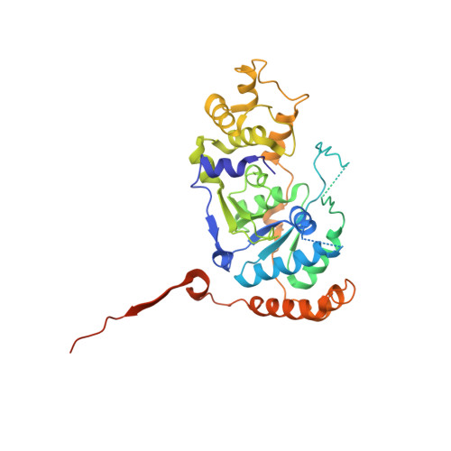
    Flap endonuclease-1 (FEN1) is a key enzyme for maintaining genomic stability and replication. Proliferating cell nuclear antigen (PCNA) binds FEN1 and stimulates its endonuclease activity. The structural basis of the FEN1-PCNA interaction was revealed by the crystal structure of the complex between human FEN1 and PCNA. The main interface involves the C-terminal tail of FEN1, which forms two beta-strands connected by a short helix, the betaA-alphaA-betaB motif, participating in beta-beta and hydrophobic interactions with PCNA. These interactions are similar to those previously observed for the p21CIP1/WAF1 peptide. However, this structure involving the full-length enzyme has revealed additional interfaces that are involved in the core domain. The interactions at the interfaces maintain the enzyme in an inactive 'locked-down' orientation and might be utilized in rapid DNA-tracking by preserving the central hole of PCNA for sliding along the DNA. A hinge region present between the core domain and the C-terminal tail of FEN1 would play a role in switching the FEN1 orientation from an inactive to an active orientation.


  • Organizational Affiliation

    Structural Biology Laboratory, Nara Institute of Science and Technology, Takayama, Ikoma, Nara, Japan.


Macromolecules
Find similar proteins by:  (by identity cutoff)  |  3D Structure
Entity ID: 1
MoleculeChains Sequence LengthOrganismDetailsImage
Flap endonuclease-1A [auth X],
B [auth Y],
C [auth Z]
379Homo sapiensMutation(s): 0 
UniProt & NIH Common Fund Data Resources
Find proteins for P39748 (Homo sapiens)
Explore P39748 
Go to UniProtKB:  P39748
PHAROS:  P39748
GTEx:  ENSG00000168496 
Entity Groups  
Sequence Clusters30% Identity50% Identity70% Identity90% Identity95% Identity100% Identity
UniProt GroupP39748
Sequence Annotations
Expand
  • Reference Sequence
Find similar proteins by:  (by identity cutoff)  |  3D Structure
Entity ID: 2
MoleculeChains Sequence LengthOrganismDetailsImage
Proliferating cell nuclear antigenD [auth A],
E [auth B],
F [auth C]
261Homo sapiensMutation(s): 0 
UniProt & NIH Common Fund Data Resources
Find proteins for P12004 (Homo sapiens)
Explore P12004 
Go to UniProtKB:  P12004
PHAROS:  P12004
GTEx:  ENSG00000132646 
Entity Groups  
Sequence Clusters30% Identity50% Identity70% Identity90% Identity95% Identity100% Identity
UniProt GroupP12004
Sequence Annotations
Expand
  • Reference Sequence
Experimental Data & Validation

Experimental Data

  • Method: X-RAY DIFFRACTION
  • Resolution: 2.90 Å
  • R-Value Free: 0.284 
  • R-Value Work: 0.220 
  • Space Group: P 21 21 21
Unit Cell:
Length ( Å )Angle ( ˚ )
a = 82.203α = 90
b = 143.383β = 90
c = 246.659γ = 90
Software Package:
Software NamePurpose
DENZOdata reduction
SCALEPACKdata scaling
MOLREPphasing
CNSrefinement

Structure Validation

View Full Validation Report


Entry History 

Deposition Data

Revision History  (Full details and data files)

  • Version 1.0: 2005-03-01
    Type: Initial release
  • Version 1.1: 2007-10-23
    Changes: Version format compliance
  • Version 1.2: 2011-07-13
    Changes: Version format compliance
  • Version 1.3: 2023-12-27
    Changes: Data collection, Database references, Derived calculations