1UJW

Structure of the complex between BtuB and Colicin E3 Receptor binding domain


Experimental Data Snapshot

  • Method: X-RAY DIFFRACTION
  • Resolution: 2.75 Å
  • R-Value Free: 0.293 
  • R-Value Work: 0.244 
  • R-Value Observed: 0.247 

wwPDB Validation   3D Report Full Report


Ligand Structure Quality Assessment 


This is version 2.1 of the entry. See complete history


Literature

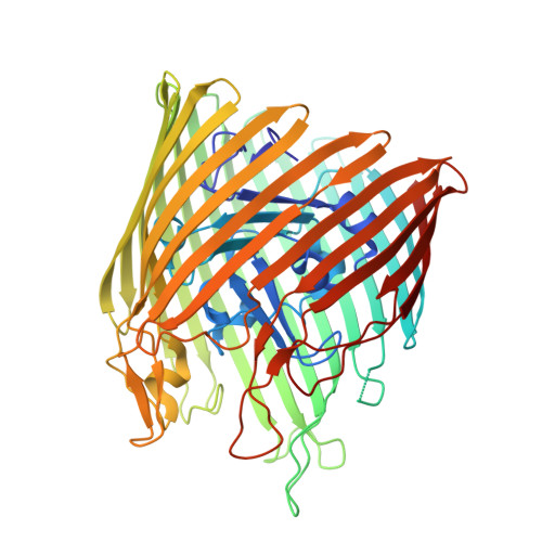
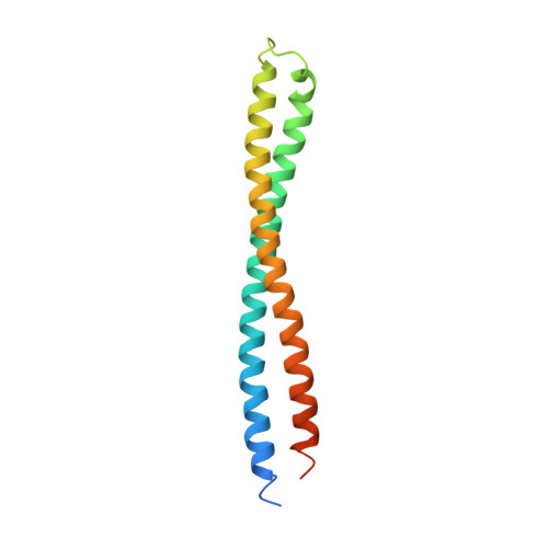
The structure of BtuB with bound colicin E3 R-domain implies a translocon

Kurisu, G.Zakharov, S.D.Zhalnina, M.V.Bano, S.Eroukova, V.Y.Rokitskaya, T.I.Antonenko, Y.N.Wiener, M.C.Cramer, W.A.

(2003) Nat Struct Biol 10: 948-954

  • DOI: https://doi.org/10.1038/nsb997
  • Primary Citation of Related Structures:  
    1UJW

  • PubMed Abstract: 

    Cellular import of colicin E3 is initiated by the Escherichia coli outer membrane cobalamin transporter, BtuB. The 135-residue 100-A coiled-coil receptor-binding domain (R135) of colicin E3 forms a 1:1 complex with BtuB whose structure at a resolution of 2.75 A is reported. Binding of R135 to the BtuB extracellular surface (DeltaG(o) = -12 kcal mol(-1)) is mediated by 27 residues of R135 near the coiled-coil apex. Formation of the R135-BtuB complex results in unfolding of R135 N- and C-terminal ends, inferred to be important for unfolding of the colicin T-domain. Small conformational changes occur in the BtuB cork and barrel domains but are insufficient to form a translocation channel. The absence of a channel and the peripheral binding of R135 imply that BtuB serves to bind the colicin, and that the coiled-coil delivers the colicin to a neighboring outer membrane protein for translocation, thus forming a colicin translocon. The translocator was concluded to be OmpF from the occlusion of OmpF channels by colicin E3.


  • Organizational Affiliation

    Department of Biological Sciences, Purdue University, Lilly Hall of Life Sciences, 915 W. State St., West Lafayette, Indiana 47907-1392, USA.


Macromolecules
Find similar proteins by:  (by identity cutoff)  |  3D Structure
Entity ID: 1
MoleculeChains Sequence LengthOrganismDetailsImage
Vitamin B12 receptor594Escherichia coliMutation(s): 0 
Membrane Entity: Yes 
UniProt
Find proteins for P06129 (Escherichia coli (strain K12))
Explore P06129 
Go to UniProtKB:  P06129
Entity Groups  
Sequence Clusters30% Identity50% Identity70% Identity90% Identity95% Identity100% Identity
UniProt GroupP06129
Sequence Annotations
Expand
  • Reference Sequence
Find similar proteins by:  (by identity cutoff)  |  3D Structure
Entity ID: 2
MoleculeChains Sequence LengthOrganismDetailsImage
Colicin E3135Escherichia coliMutation(s): 0 
UniProt
Find proteins for P00646 (Escherichia coli)
Explore P00646 
Go to UniProtKB:  P00646
Entity Groups  
Sequence Clusters30% Identity50% Identity70% Identity90% Identity95% Identity100% Identity
UniProt GroupP00646
Sequence Annotations
Expand
  • Reference Sequence
Small Molecules
Ligands 5 Unique
IDChains Name / Formula / InChI Key2D Diagram3D Interactions
GP1
Query on GP1

Download Ideal Coordinates CCD File 
C [auth A],
D [auth A]
2-amino-2-deoxy-1-O-phosphono-alpha-D-glucopyranose
C6 H14 N O8 P
YMJBYRVFGYXULK-QZABAPFNSA-N
LIM
Query on LIM

Download Ideal Coordinates CCD File 
E [auth A],
N [auth A]
3-OXO-PENTADECANOIC ACID
C15 H28 O3
CJTNJZWHYGHVCD-UHFFFAOYSA-N
LDA
Query on LDA

Download Ideal Coordinates CCD File 
F [auth A]
G [auth A]
H [auth A]
I [auth A]
J [auth A]
F [auth A],
G [auth A],
H [auth A],
I [auth A],
J [auth A],
K [auth A],
L [auth A]
LAURYL DIMETHYLAMINE-N-OXIDE
C14 H31 N O
SYELZBGXAIXKHU-UHFFFAOYSA-N
AAE
Query on AAE

Download Ideal Coordinates CCD File 
M [auth A]ACETOACETIC ACID
C4 H6 O3
WDJHALXBUFZDSR-UHFFFAOYSA-N
GOL
Query on GOL

Download Ideal Coordinates CCD File 
O [auth A]
P [auth A]
Q [auth A]
R [auth A]
S [auth A]
O [auth A],
P [auth A],
Q [auth A],
R [auth A],
S [auth A],
T [auth A],
U [auth A],
V [auth A]
GLYCEROL
C3 H8 O3
PEDCQBHIVMGVHV-UHFFFAOYSA-N
Experimental Data & Validation

Experimental Data

  • Method: X-RAY DIFFRACTION
  • Resolution: 2.75 Å
  • R-Value Free: 0.293 
  • R-Value Work: 0.244 
  • R-Value Observed: 0.247 
  • Space Group: P 21 21 21
Unit Cell:
Length ( Å )Angle ( ˚ )
a = 76.933α = 90
b = 80.098β = 90
c = 233.595γ = 90
Software Package:
Software NamePurpose
REFMACrefinement
HKL-2000data reduction
HKL-2000data scaling
AMoREphasing

Structure Validation

View Full Validation Report


Ligand Structure Quality Assessment 


Entry History 

Deposition Data

Revision History  (Full details and data files)

  • Version 1.0: 2003-11-25
    Type: Initial release
  • Version 1.1: 2008-04-27
    Changes: Version format compliance
  • Version 1.2: 2011-07-13
    Changes: Advisory, Version format compliance
  • Version 2.0: 2020-07-29
    Type: Remediation
    Reason: Carbohydrate remediation
    Changes: Atomic model, Data collection, Derived calculations, Structure summary
  • Version 2.1: 2023-10-25
    Changes: Data collection, Database references, Derived calculations, Refinement description, Structure summary