1S4Y

Crystal structure of the activin/actrIIb extracellular domain


Experimental Data Snapshot

  • Method: X-RAY DIFFRACTION
  • Resolution: 2.30 Å
  • R-Value Free: 0.286 
  • R-Value Work: 0.206 
  • R-Value Observed: 0.210 

wwPDB Validation   3D Report Full Report


This is version 1.2 of the entry. See complete history


Literature

A Flexible Activin Explains the Membrane-Dependent Cooperative Assembly of TGF-beta Family Receptors.

Greenwald, J.Vega, M.E.Allendorph, G.P.Fischer, W.H.Vale, W.Choe, S.

(2004) Mol Cell 15: 485-489

  • DOI: https://doi.org/10.1016/j.molcel.2004.07.011
  • Primary Citation of Related Structures:  
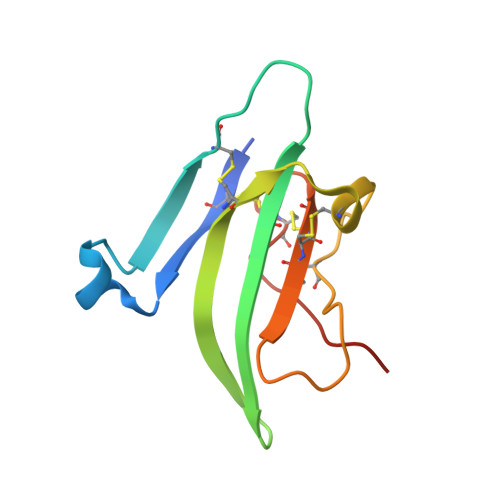
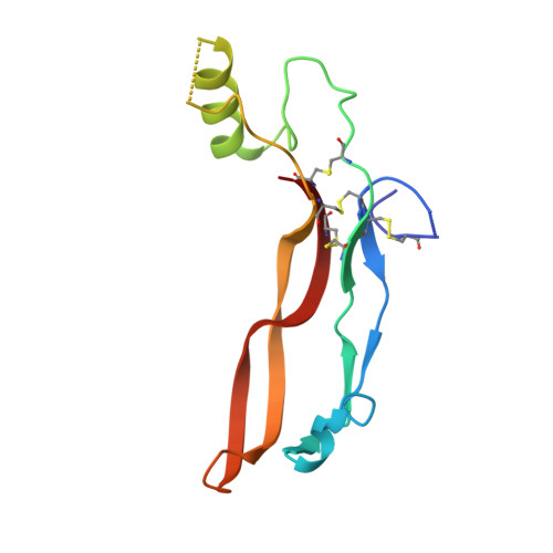
    1S4Y

  • PubMed Abstract: 

    A new crystal structure of activin in complex with the extracellular domain of its type II receptor (ActRIIb-ECD) shows that the ligand exhibits an unexpected flexibility. The motion in the activin dimer disrupts its type I receptor interface, which may account for the disparity in its affinity for type I versus type II receptors. We have measured the affinities of activin and its antagonist inhibin for ActRIIb-ECD and found that the affinity of the 2-fold symmetric homodimer activin for ActRIIb-ECD depends on the availability of two spatially coupled ActRIIb-ECD molecules, whereas the affinity of the heterodimer inhibin does not. Our results indicate that activin's affinity for its two receptor types is greatly influenced by their membrane-restricted setting. We propose that activin affinity is modulated by the ligand flexibility and that cooperativity is achieved by binding to two ActRII chains that immobilize activin in a type I binding-competent orientation.


  • Organizational Affiliation

    Structural Biology Laboratory, The Salk Institute, La Jolla, CA 92037, USA.


Macromolecules
Find similar proteins by:  (by identity cutoff)  |  3D Structure
Entity ID: 1
MoleculeChains Sequence LengthOrganismDetailsImage
Activin receptor type IIB precursor
A, C
98Mus musculusMutation(s): 0 
Gene Names: ACVR2B
EC: 2.7.1.37
UniProt & NIH Common Fund Data Resources
Find proteins for P27040 (Mus musculus)
Explore P27040 
Go to UniProtKB:  P27040
IMPC:  MGI:87912
Entity Groups  
Sequence Clusters30% Identity50% Identity70% Identity90% Identity95% Identity100% Identity
UniProt GroupP27040
Sequence Annotations
Expand
  • Reference Sequence
Find similar proteins by:  (by identity cutoff)  |  3D Structure
Entity ID: 2
MoleculeChains Sequence LengthOrganismDetailsImage
Inhibin beta A chain
B, D
116Homo sapiensMutation(s): 0 
Gene Names: INHBA
UniProt & NIH Common Fund Data Resources
Find proteins for P08476 (Homo sapiens)
Explore P08476 
Go to UniProtKB:  P08476
PHAROS:  P08476
GTEx:  ENSG00000122641 
Entity Groups  
Sequence Clusters30% Identity50% Identity70% Identity90% Identity95% Identity100% Identity
UniProt GroupP08476
Sequence Annotations
Expand
  • Reference Sequence
Experimental Data & Validation

Experimental Data

  • Method: X-RAY DIFFRACTION
  • Resolution: 2.30 Å
  • R-Value Free: 0.286 
  • R-Value Work: 0.206 
  • R-Value Observed: 0.210 
  • Space Group: P 21 21 2
Unit Cell:
Length ( Å )Angle ( ˚ )
a = 86.29α = 90
b = 120.627β = 90
c = 43.55γ = 90
Software Package:
Software NamePurpose
REFMACrefinement
HKL-2000data reduction
SCALEPACKdata scaling
EPMRphasing

Structure Validation

View Full Validation Report


Entry History 

Deposition Data

Revision History  (Full details and data files)

  • Version 1.0: 2004-08-10
    Type: Initial release
  • Version 1.1: 2008-04-29
    Changes: Version format compliance
  • Version 1.2: 2011-07-13
    Changes: Advisory, Version format compliance