Solution Structure and Backbone Dynamics of the Non-receptor Protein-tyrosine Kinase-6 Src Homology 2 Domain
Hong, E., Shin, J., Kim, H.I., Lee, S.T., Lee, W.(2004) J Biol Chem 279: 29700-29708
- PubMed: 15056653
- DOI: https://doi.org/10.1074/jbc.M313185200
- Primary Citation of Related Structures:
1RJA - PubMed Abstract:
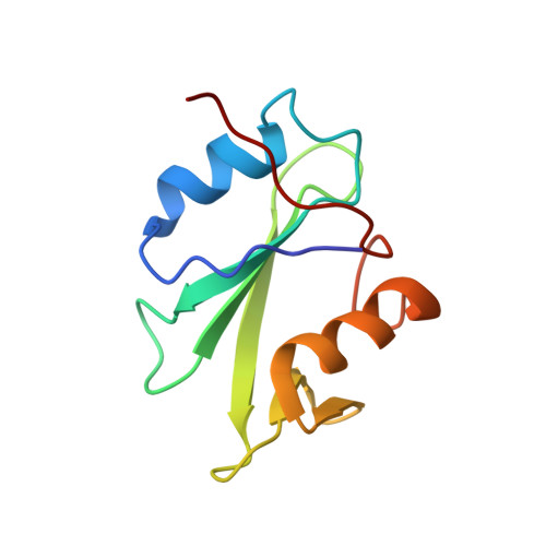
Human protein-tyrosine kinase-6 (PTK6, also known as breast tumor kinase (Brk)) is a member of the non-receptor protein-tyrosine kinase family and is expressed in two-thirds of all breast tumors. To understand the structural basis of PTK6 function, we have determined the solution structure and backbone dynamics of the PTK6-Src homology 2 (SH2) domain using multidimensional NMR spectroscopy. The solution structure clearly indicates that the SH2 domain of human PTK6 contains a consensus alpha/beta-fold and a Tyr(P) peptide binding surface, which are common to other SH2 domains. However, two of the alpha-helices (alphaA and alphaB) are located on opposite faces of the central beta-sheet. In addition, the topological arrangement of a central four-stranded antiparallel beta-sheet (strands betaA, betaB, betaC, and betaD) differs from that of other Src family members. Backbone dynamics and Tyr(P) peptide titration experiments revealed that the putative ligand binding sites of the PTK6-SH2 domain undergo distinctive internal motions when compared with other regions of the protein. Surface plasmon resonance analysis showed that the Tyr(P) peptide had a dissociation constant of about 60 microm, which is substantially weaker binding than previously reported for Src family members. The solution structure together with data from the ligand binding mode of PTK6-SH2 provides insight into the molecular basis of the autoinhibitory role of PTK6.
Organizational Affiliation:
Department of Biochemistry and Protein Network Research Center, College of Science, Yonsei University, Seoul 120-749, Korea.